 Movie
Movie Controller


+ Open data

- Basic information
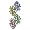
| Entry | Database: PDB / ID: 7bvy | ||||||
|---|---|---|---|---|---|---|---|
| Title | Crystal structure of MreB 5 of Spiroplasma citri bound to AMPPNP | ||||||
 Components Components | Cell shape determining protein MreB | ||||||
 Keywords Keywords | PROTEIN FIBRIL / ATPase | ||||||
| Function / homology |  Function and homology information Function and homology information | ||||||
| Biological species |  Spiroplasma citri (bacteria) Spiroplasma citri (bacteria) | ||||||
| Method |  X-RAY DIFFRACTION / SYNCHROTRON / MOLECULAR REPLACEMENT / molecular replacement / Resolution: 2.5 Å X-RAY DIFFRACTION / SYNCHROTRON / MOLECULAR REPLACEMENT / molecular replacement / Resolution: 2.5 Å | ||||||
 Authors Authors | Pande, V. / Gayathri, P. | ||||||
| Funding support |  India, 1items India, 1items
| ||||||
 Citation Citation | Journal: Curr.Biol. / Year: 2020 Title: MreB5 Is a Determinant of Rod-to-Helical Transition in the Cell-Wall-less Bacterium Spiroplasma. Authors: Harne, S. / Duret, S. / Pande, V. / Bapat, M. / Beven, L. / Gayathri, P. | ||||||
| History |
|
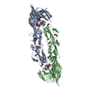
- Structure visualization
| Structure viewer | Molecule: MolmilJmol/JSmol |
|---|
- Downloads & links
-Download
| PDBx/mmCIF format | 7bvy.cif.gz | 84.2 KB | Display | PDBx/mmCIF format |
|---|---|---|---|---|
| PDB format | pdb7bvy.ent.gz | 59 KB | Display | PDB format |
| PDBx/mmJSON format | 7bvy.json.gz | Tree view | PDBx/mmJSON format | |
| Others |  Other downloads Other downloads |
-Validation report
| Arichive directory | https://data.pdbj.org/pub/pdb/validation_reports/bv/7bvyftp://data.pdbj.org/pub/pdb/validation_reports/bv/7bvy | HTTPS FTP |
|---|
-Related structure data
| Related structure data | 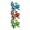 7bvzSC S: Starting model for refinement C: citing same article ( |
|---|---|
| Similar structure data |


-Links
 PDBj
PDBj


- Assembly
| Deposited unit | 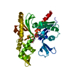
| ||||||||
|---|---|---|---|---|---|---|---|---|---|
| 1 | 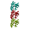
| ||||||||
| Unit cell |
|
-Components
-Protein , 1 types, 1 molecules A
| #1: Protein | Mass: 39669.770 Da / Num. of mol.: 1 Source method: isolated from a genetically manipulated source Details: cell wall less bacteria / Source: (gene. exp.) Spiroplasma citri (bacteria) / Gene: mreB5, FRX96_09810, SCITRI_001914, SPICI01A_049 / Variant: GII3 / Plasmid: pHis17 / Production host: |
|---|
-Non-polymers , 5 types, 113 molecules 


| #2: Chemical | ChemComp-ANP /  Mass: 506.196 Da / Num. of mol.: 1 / Source method: obtained synthetically / Formula: C10H17N6O12P3 / Comment: AMP-PNP, energy-carrying molecule analogue*YM Mass: 506.196 Da / Num. of mol.: 1 / Source method: obtained synthetically / Formula: C10H17N6O12P3 / Comment: AMP-PNP, energy-carrying molecule analogue*YM |
|---|---|
| #3: Chemical | ChemComp-PO4 /  Mass: 94.971 Da / Num. of mol.: 1 / Source method: obtained synthetically / Formula: PO4 Mass: 94.971 Da / Num. of mol.: 1 / Source method: obtained synthetically / Formula: PO4 |
| #4: Chemical | ChemComp-MG /  Mass: 24.305 Da / Num. of mol.: 1 / Source method: obtained synthetically / Formula: Mg Mass: 24.305 Da / Num. of mol.: 1 / Source method: obtained synthetically / Formula: Mg |
| #5: Chemical | ChemComp-K /  Mass: 39.098 Da / Num. of mol.: 1 / Source method: obtained synthetically / Formula: K Mass: 39.098 Da / Num. of mol.: 1 / Source method: obtained synthetically / Formula: K |
| #6: Water | ChemComp-HOH / Mass: 18.015 Da / Num. of mol.: 109 / Source method: isolated from a natural source / Formula: H2O |
-Details
| Has ligand of interest | N |
|---|
-Experimental details
-Experiment
| Experiment | Method: X-RAY DIFFRACTION / Number of used crystals: 1 |
|---|
- Sample preparation
| Crystal | Density Matthews: 2.39 Å3/Da / Density % sol: 48.62 % / Description: Thin Needle- like crystals in a bunch |
|---|---|
| Crystal grow | Temperature: 291 K / Method: vapor diffusion, sitting drop / pH: 7.8 Details: 0.15M Na-K phosphate, 16% PEG 3350, pH 7.8, 2mM AMPPNP, 2mM MgCl2 |
-Data collection
| Diffraction | Mean temperature: 100 K / Serial crystal experiment: N |
|---|---|
| Diffraction source | Source: SYNCHROTRON / Site: ESRF  / Beamline: MASSIF-3 / Wavelength: 0.9677 Å / Beamline: MASSIF-3 / Wavelength: 0.9677 Å |
| Detector | Type: DECTRIS EIGER X 4M / Detector: PIXEL / Date: Oct 1, 2018 |
| Radiation | Protocol: SINGLE WAVELENGTH / Monochromatic (M) / Laue (L): M / Scattering type: x-ray |
| Radiation wavelength | Wavelength: 0.9677 Å / Relative weight: 1 |
| Reflection | Resolution: 2.5→49.838 Å / Num. obs: 13181 / % possible obs: 98.9 % / Redundancy: 6.9 % / Biso Wilson estimate: 26.47 Å2 / CC1/2: 0.99 / Rmerge(I) obs: 0.2 / Rpim(I) all: 0.12 / Net I/σ(I): 7 |
| Reflection shell | Resolution: 2.5→2.6 Å / Redundancy: 7.1 % / Rmerge(I) obs: 0.89 / Mean I/σ(I) obs: 2.2 / Num. unique obs: 1461 / CC1/2: 0.82 / Rpim(I) all: 0.54 / % possible all: 98.3 |
-Phasing
| Phasing | Method: molecular replacement |
|---|
- Processing
| Software |
| ||||||||||||||||||||||||||||||||||||||||||||||||||||||||||||
|---|---|---|---|---|---|---|---|---|---|---|---|---|---|---|---|---|---|---|---|---|---|---|---|---|---|---|---|---|---|---|---|---|---|---|---|---|---|---|---|---|---|---|---|---|---|---|---|---|---|---|---|---|---|---|---|---|---|---|---|---|---|
| Refinement | Method to determine structure: MOLECULAR REPLACEMENT Starting model: 7BVZ Resolution: 2.5→49.838 Å / SU ML: 0.32 / Cross valid method: THROUGHOUT / σ(F): 0.26 / Phase error: 26.62
| ||||||||||||||||||||||||||||||||||||||||||||||||||||||||||||
| Solvent computation | Shrinkage radii: 0.9 Å / VDW probe radii: 1.11 Å | ||||||||||||||||||||||||||||||||||||||||||||||||||||||||||||
| Displacement parameters | Biso max: 76.22 Å2 / Biso mean: 32.1149 Å2 / Biso min: 14.34 Å2 | ||||||||||||||||||||||||||||||||||||||||||||||||||||||||||||
| Refinement step | Cycle: final / Resolution: 2.5→49.838 Å
| ||||||||||||||||||||||||||||||||||||||||||||||||||||||||||||
| Refine LS restraints |
| ||||||||||||||||||||||||||||||||||||||||||||||||||||||||||||
| LS refinement shell | Refine-ID: X-RAY DIFFRACTION / Rfactor Rfree error: 0
|
