 Movie
Movie Controller

[English] 日本語
 Yorodumi
- PDB-4m00: Crystal structure of the ligand binding region of staphylococcal ... -
+ Open data

- Basic information
| Entry | Database: PDB / ID: 4m00 | ||||||||||||
|---|---|---|---|---|---|---|---|---|---|---|---|---|---|
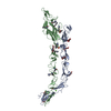
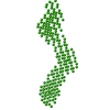
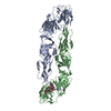
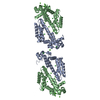
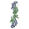
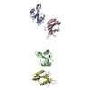
| Title | Crystal structure of the ligand binding region of staphylococcal adhesion SraP | ||||||||||||
 Components Components | Serine-rich adhesin for platelets | ||||||||||||
 Keywords Keywords | CELL ADHESION / All Beta / Adhesion / Carbohydrate/Sugar Binding | ||||||||||||
| Function / homology |  Function and homology information Function and homology information | ||||||||||||
| Biological species |   Staphylococcus aureus (bacteria) Staphylococcus aureus (bacteria) | ||||||||||||
| Method |  X-RAY DIFFRACTION / SYNCHROTRON / MOLECULAR REPLACEMENT / Resolution: 2.05 Å X-RAY DIFFRACTION / SYNCHROTRON / MOLECULAR REPLACEMENT / Resolution: 2.05 Å | ||||||||||||
 Authors Authors | Yang, Y.H. / Jiang, Y.L. / Zhang, J. / Wang, L. / Chen, Y. / Zhou, C.Z. | ||||||||||||
 Citation Citation | Journal: Plos Pathog. / Year: 2014 Title: Structural Insights into SraP-Mediated Staphylococcus aureus Adhesion to Host Cells Authors: Yang, Y.H. / Jiang, Y.L. / Zhang, J. / Wang, L. / Bai, X.H. / Zhang, S.J. / Ren, Y.M. / Li, N. / Zhang, Y.H. / Zhang, Z. / Gong, Q. / Mei, Y. / Xue, T. / Zhang, J.R. / Chen, Y. / Zhou, C.Z. | ||||||||||||
| History |
|
- Structure visualization
| Structure viewer | Molecule: MolmilJmol/JSmol |
|---|
- Downloads & links
-Download
| PDBx/mmCIF format | 4m00.cif.gz | 125.8 KB | Display | PDBx/mmCIF format |
|---|---|---|---|---|
| PDB format | pdb4m00.ent.gz | 92.2 KB | Display | PDB format |
| PDBx/mmJSON format | 4m00.json.gz | Tree view | PDBx/mmJSON format | |
| Others |  Other downloads Other downloads |
-Validation report
| Arichive directory | https://data.pdbj.org/pub/pdb/validation_reports/m0/4m00ftp://data.pdbj.org/pub/pdb/validation_reports/m0/4m00 | HTTPS FTP |
|---|
-Related structure data
| Related structure data | 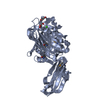 4m01SC 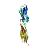 4m02C  4m03C S: Starting model for refinement C: citing same article ( |
|---|---|
| Similar structure data |


-Links
 PDBj
PDBj

- Assembly
| Deposited unit | 
| ||||||||
|---|---|---|---|---|---|---|---|---|---|
| 1 |
| ||||||||
| Unit cell |
|
-Components
| #1: Protein | Mass: 56361.020 Da / Num. of mol.: 1 / Fragment: ligand binding region, UNP residues 245-751 Source method: isolated from a genetically manipulated source Source: (gene. exp.) Staphylococcus aureus (bacteria) / Strain: NCTC 8325 / Gene: sraP / Plasmid: pET28a / Production host: | ||||||
|---|---|---|---|---|---|---|---|
| #2: Polysaccharide | beta-D-fructofuranose-(2-1)-alpha-D-glucopyranose / sucrose  Source method: isolated from a genetically manipulated source Details: oligosaccharide with reducing-end-to-reducing-end glycosidic bond References: sucrose | ||||||
| #3: Chemical |   Mass: 40.078 Da / Num. of mol.: 3 / Source method: obtained synthetically / Formula: Ca Mass: 40.078 Da / Num. of mol.: 3 / Source method: obtained synthetically / Formula: Ca#4: Chemical | ChemComp-MES / |   Mass: 195.237 Da / Num. of mol.: 1 / Source method: obtained synthetically / Formula: C6H13NO4S / Comment: pH buffer*YM Mass: 195.237 Da / Num. of mol.: 1 / Source method: obtained synthetically / Formula: C6H13NO4S / Comment: pH buffer*YM#5: Water | ChemComp-HOH / |  Mass: 18.015 Da / Num. of mol.: 618 / Source method: isolated from a natural source / Formula: H2O Mass: 18.015 Da / Num. of mol.: 618 / Source method: isolated from a natural source / Formula: H2OHas protein modification | N | |
-Experimental details
-Experiment
| Experiment | Method: X-RAY DIFFRACTION / Number of used crystals: 1 |
|---|
- Sample preparation
| Crystal | Density Matthews: 4.87 Å3/Da / Density % sol: 74.72 % |
|---|---|
| Crystal grow | Temperature: 301 K / Method: vapor diffusion, hanging drop / pH: 6 Details: 0.8M ammonium sulfate, 0.1M MES, pH 6.0, VAPOR DIFFUSION, HANGING DROP, temperature 301K |
-Data collection
| Diffraction | Mean temperature: 100 K |
|---|---|
| Diffraction source | Source: SYNCHROTRON / Site: SSRF  / Beamline: BL17U / Wavelength: 0.9793 Å / Beamline: BL17U / Wavelength: 0.9793 Å |
| Detector | Type: ADSC QUANTUM 315r / Detector: CCD / Date: Apr 16, 2013 |
| Radiation | Monochromator: Si 111 / Protocol: SINGLE WAVELENGTH / Monochromatic (M) / Laue (L): M / Scattering type: x-ray |
| Radiation wavelength | Wavelength: 0.9793 Å / Relative weight: 1 |
| Reflection | Resolution: 2.05→50 Å / Num. all: 69226 / Num. obs: 69226 / % possible obs: 100 % / Observed criterion σ(F): -3 / Observed criterion σ(I): 0 / Redundancy: 6.9 % / Rmerge(I) obs: 0.137 / Rsym value: 0.137 / Net I/σ(I): 13.8 |
| Reflection shell | Resolution: 2.05→2.12 Å / Redundancy: 6.8 % / Rmerge(I) obs: 0.385 / Mean I/σ(I) obs: 5.754 / Num. unique all: 475054 / Rsym value: 0.385 / % possible all: 100 |
- Processing
| Software |
| |||||||||||||||||||||||||||||||||||||||||||||
|---|---|---|---|---|---|---|---|---|---|---|---|---|---|---|---|---|---|---|---|---|---|---|---|---|---|---|---|---|---|---|---|---|---|---|---|---|---|---|---|---|---|---|---|---|---|---|
| Refinement | Method to determine structure: MOLECULAR REPLACEMENT Starting model: 4M01 Resolution: 2.05→41.54 Å / Cor.coef. Fo:Fc: 0.951 / Cor.coef. Fo:Fc free: 0.939 / SU B: 2.558 / SU ML: 0.07 / Cross valid method: THROUGHOUT / ESU R: 0.113 / ESU R Free: 0.11 / Stereochemistry target values: MAXIMUM LIKELIHOOD / Details: HYDROGENS HAVE BEEN USED IF PRESENT IN THE INPUT
| |||||||||||||||||||||||||||||||||||||||||||||
| Solvent computation | Ion probe radii: 0.8 Å / Shrinkage radii: 0.8 Å / VDW probe radii: 1.2 Å / Solvent model: MASK | |||||||||||||||||||||||||||||||||||||||||||||
| Displacement parameters | Biso mean: 29.548 Å2
| |||||||||||||||||||||||||||||||||||||||||||||
| Refinement step | Cycle: LAST / Resolution: 2.05→41.54 Å
| |||||||||||||||||||||||||||||||||||||||||||||
| Refine LS restraints |
| |||||||||||||||||||||||||||||||||||||||||||||
| LS refinement shell | Resolution: 2.05→2.103 Å / Total num. of bins used: 20
|
