 Movie
Movie Controller

[English] 日本語
 Yorodumi
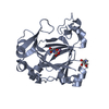
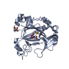
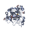
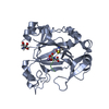
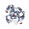
- PDB-7dyx: Human JMJD5 in complex with MN and 5-((2-cyclopropylbenzyl)amino)... -
+ Open data

- Basic information
| Entry | Database: PDB / ID: 7dyx | ||||||
|---|---|---|---|---|---|---|---|
| Title | Human JMJD5 in complex with MN and 5-((2-cyclopropylbenzyl)amino)pyridine-2,4-dicarboxylic acid. | ||||||
 Components Components | Bifunctional peptidase and arginyl-hydroxylase JMJD5 | ||||||
 Keywords Keywords | OXIDOREDUCTASE / JmjC domain-containing protein 5 / JMJD5 / dioxygenase | ||||||
| Function / homology |  Function and homology information Function and homology information[protein]-arginine 3-hydroxylase / peptidyl-arginine 3-dioxygenase activity / histone H3K36 demethylase activity / Hydrolases; Acting on peptide bonds (peptidases) / Protein hydroxylation / aminopeptidase activity / regulation of signal transduction by p53 class mediator / circadian regulation of gene expression / protein destabilization / G2/M transition of mitotic cell cycle ...[protein]-arginine 3-hydroxylase / peptidyl-arginine 3-dioxygenase activity / histone H3K36 demethylase activity / Hydrolases; Acting on peptide bonds (peptidases) / Protein hydroxylation / aminopeptidase activity / regulation of signal transduction by p53 class mediator / circadian regulation of gene expression / protein destabilization / G2/M transition of mitotic cell cycle / p53 binding / chromosome / fibroblast proliferation / endopeptidase activity / in utero embryonic development / negative regulation of DNA-templated transcription / chromatin binding / positive regulation of DNA-templated transcription / proteolysis / nucleoplasm / metal ion binding / nucleus / cytosol Similarity search - Function | ||||||
| Biological species |  Homo sapiens (human) Homo sapiens (human) | ||||||
| Method |  X-RAY DIFFRACTION / SYNCHROTRON / MOLECULAR REPLACEMENT / Resolution: 2.27 Å X-RAY DIFFRACTION / SYNCHROTRON / MOLECULAR REPLACEMENT / Resolution: 2.27 Å | ||||||
 Authors Authors | Nakashima, Y. / Brewitz, L. / Schofield, C.J. | ||||||
 Citation Citation | Journal: J.Med.Chem. / Year: 2023 Title: 5-Substituted Pyridine-2,4-dicarboxylate Derivatives Have Potential for Selective Inhibition of Human Jumonji-C Domain-Containing Protein 5. Authors: Brewitz, L. / Nakashima, Y. / Piasecka, S.K. / Salah, E. / Fletcher, S.C. / Tumber, A. / Corner, T.P. / Kennedy, T.J. / Fiorini, G. / Thalhammer, A. / Christensen, K.E. / Coleman, M.L. / Schofield, C.J. | ||||||
| History |
|
- Structure visualization
| Structure viewer | Molecule: MolmilJmol/JSmol |
|---|
- Downloads & links
-Download
| PDBx/mmCIF format | 7dyx.cif.gz | 191.6 KB | Display | PDBx/mmCIF format |
|---|---|---|---|---|
| PDB format | pdb7dyx.ent.gz | 127.4 KB | Display | PDB format |
| PDBx/mmJSON format | 7dyx.json.gz | Tree view | PDBx/mmJSON format | |
| Others |  Other downloads Other downloads |
-Validation report
| Arichive directory | https://data.pdbj.org/pub/pdb/validation_reports/dy/7dyxftp://data.pdbj.org/pub/pdb/validation_reports/dy/7dyx | HTTPS FTP |
|---|
-Related structure data
| Related structure data | 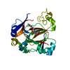 7dytC  7dyuC  7dyvC 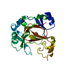 7dywC  6f4pS S: Starting model for refinement C: citing same article ( |
|---|---|
| Similar structure data |

-Links
 PDBj
PDBj
- Assembly
| Deposited unit | 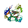
| ||||||||||||
|---|---|---|---|---|---|---|---|---|---|---|---|---|---|
| 1 |
| ||||||||||||
| Unit cell |
|
-Components
| #1: Protein | Mass: 27261.873 Da / Num. of mol.: 1 Source method: isolated from a genetically manipulated source Source: (gene. exp.) Homo sapiens (human) / Gene: KDM8, JMJD5 / Production host:  References: UniProt: Q8N371, Oxidoreductases; Acting on paired donors, with incorporation or reduction of molecular oxygen; With 2-oxoglutarate as one donor, and incorporation of one atom of oxygen ...References: UniProt: Q8N371, Oxidoreductases; Acting on paired donors, with incorporation or reduction of molecular oxygen; With 2-oxoglutarate as one donor, and incorporation of one atom of oxygen into each donor, Hydrolases; Acting on peptide bonds (peptidases) |
|---|---|
| #2: Chemical | ChemComp-MN /   Mass: 54.938 Da / Num. of mol.: 1 / Source method: obtained synthetically / Formula: Mn Mass: 54.938 Da / Num. of mol.: 1 / Source method: obtained synthetically / Formula: Mn |
| #3: Chemical | ChemComp-HRR /   Mass: 312.320 Da / Num. of mol.: 1 / Source method: obtained synthetically / Formula: C17H16N2O4 / Feature type: SUBJECT OF INVESTIGATION Mass: 312.320 Da / Num. of mol.: 1 / Source method: obtained synthetically / Formula: C17H16N2O4 / Feature type: SUBJECT OF INVESTIGATION |
| #4: Water | ChemComp-HOH /  Mass: 18.015 Da / Num. of mol.: 57 / Source method: isolated from a natural source / Formula: H2O Mass: 18.015 Da / Num. of mol.: 57 / Source method: isolated from a natural source / Formula: H2O |
| Has ligand of interest | Y |
-Experimental details
-Experiment
| Experiment | Method: X-RAY DIFFRACTION / Number of used crystals: 1 |
|---|
- Sample preparation
| Crystal | Density Matthews: 2.28 Å3/Da / Density % sol: 46.16 % |
|---|---|
| Crystal grow | Temperature: 277 K / Method: vapor diffusion, sitting drop / pH: 7.5 Details: 100mM HEPES sodium, 200mM magnesium chloride hexahydrate, 25% w/v PEG 3350, 1mM manganese chloride |
-Data collection
| Diffraction | Mean temperature: 100 K / Serial crystal experiment: N |
|---|---|
| Diffraction source | Source: SYNCHROTRON / Site: ESRF  / Beamline: MASSIF-1 / Wavelength: 0.965459 Å / Beamline: MASSIF-1 / Wavelength: 0.965459 Å |
| Detector | Type: DECTRIS PILATUS3 2M / Detector: PIXEL / Date: Nov 3, 2020 |
| Radiation | Protocol: SINGLE WAVELENGTH / Monochromatic (M) / Laue (L): M / Scattering type: x-ray |
| Radiation wavelength | Wavelength: 0.965459 Å / Relative weight: 1 |
| Reflection | Resolution: 2.27→48.92 Å / Num. obs: 21676 / % possible obs: 97.1 % / Redundancy: 2.32 % / Biso Wilson estimate: 38.22 Å2 / CC1/2: 0.943 / Rmerge(I) obs: 0.435 / Net I/σ(I): 2.69 |
| Reflection shell | Resolution: 2.27→2.41 Å / Rmerge(I) obs: 2.966 / Mean I/σ(I) obs: 0.35 / Num. unique obs: 215 / CC1/2: 0.098 / % possible all: 98.2 |
- Processing
| Software |
| |||||||||||||||||||||||||||||||||||||||||||||||||||||||||||||||||||||||||||
|---|---|---|---|---|---|---|---|---|---|---|---|---|---|---|---|---|---|---|---|---|---|---|---|---|---|---|---|---|---|---|---|---|---|---|---|---|---|---|---|---|---|---|---|---|---|---|---|---|---|---|---|---|---|---|---|---|---|---|---|---|---|---|---|---|---|---|---|---|---|---|---|---|---|---|---|---|
| Refinement | Method to determine structure: MOLECULAR REPLACEMENT Starting model: 6F4P Resolution: 2.27→41.54 Å / SU ML: 0.3556 / Cross valid method: FREE R-VALUE / σ(F): 0.97 / Phase error: 29.117 Stereochemistry target values: GeoStd + Monomer Library + CDL v1.2
| |||||||||||||||||||||||||||||||||||||||||||||||||||||||||||||||||||||||||||
| Solvent computation | Shrinkage radii: 0.9 Å / VDW probe radii: 1.11 Å / Solvent model: FLAT BULK SOLVENT MODEL | |||||||||||||||||||||||||||||||||||||||||||||||||||||||||||||||||||||||||||
| Displacement parameters | Biso mean: 48.82 Å2 | |||||||||||||||||||||||||||||||||||||||||||||||||||||||||||||||||||||||||||
| Refinement step | Cycle: LAST / Resolution: 2.27→41.54 Å
| |||||||||||||||||||||||||||||||||||||||||||||||||||||||||||||||||||||||||||
| Refine LS restraints |
| |||||||||||||||||||||||||||||||||||||||||||||||||||||||||||||||||||||||||||
| LS refinement shell |
| |||||||||||||||||||||||||||||||||||||||||||||||||||||||||||||||||||||||||||
| Refinement TLS params. | Method: refined / Refine-ID: X-RAY DIFFRACTION
| |||||||||||||||||||||||||||||||||||||||||||||||||||||||||||||||||||||||||||
| Refinement TLS group |
|
