[English] 日本語
Yorodumi
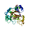
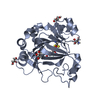
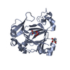
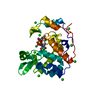
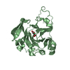
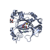
- PDB-5fbj: Complex structure of JMJD5 and substrate -

+
Open data


ID or keywords:

Loading...

-
Basic information

Entry
Database: PDB / ID: 5fbj
TitleComplex structure of JMJD5 and substrate
ComponentsLysine-specific demethylase 8
KeywordsOXIDOREDUCTASE / Histone enzyme
Function / homology
Function and homology information


[protein]-arginine 3-hydroxylase / peptidyl-arginine 3-dioxygenase activity / histone H3K36 demethylase activity / Hydrolases; Acting on peptide bonds (peptidases) / Protein hydroxylation / aminopeptidase activity / methylated histone binding / regulation of signal transduction by p53 class mediator / circadian regulation of gene expression / protein destabilization ...[protein]-arginine 3-hydroxylase / peptidyl-arginine 3-dioxygenase activity / histone H3K36 demethylase activity / Hydrolases; Acting on peptide bonds (peptidases) / Protein hydroxylation / aminopeptidase activity / methylated histone binding / regulation of signal transduction by p53 class mediator / circadian regulation of gene expression / protein destabilization / G2/M transition of mitotic cell cycle / p53 binding / chromosome / fibroblast proliferation / endopeptidase activity / in utero embryonic development / negative regulation of DNA-templated transcription / chromatin binding / positive regulation of DNA-templated transcription / proteolysis / nucleoplasm / metal ion binding / nucleus / cytosol
Similarity search - Function
Cupin-like domain 8 / Cupin-like domain / Cupin / A domain family that is part of the cupin metalloenzyme superfamily. / JmjC domain / JmjC domain profile. / Jelly Rolls / Sandwich / Mainly Beta
Similarity search - Domain/homology
2-OXOGLUTARIC ACID / Chem-NMM / Bifunctional peptidase and arginyl-hydroxylase JMJD5
Similarity search - Component
Biological speciesHomo sapiens (human)
MethodX-RAY DIFFRACTION / MOLECULAR REPLACEMENT / Resolution: 2.42 Å
AuthorsLiu, H.L. / Wang, Y. / Wang, C. / Zhang, G.Y.
CitationJournal: To Be Published
Title: to be published
Authors: Liu, H.L. / Wang, Y. / Wang, C. / Dai, S.D. / Zhang, G.Y.
History
DepositionDec 14, 2015Deposition site: RCSB / Processing site: RCSB
Revision 1.0Dec 14, 2016Provider: repository / Type: Initial release
Revision 1.1Sep 27, 2023Group: Advisory / Data collection ...Advisory / Data collection / Database references / Refinement description
Category: chem_comp_atom / chem_comp_bond ...chem_comp_atom / chem_comp_bond / database_2 / pdbx_initial_refinement_model / pdbx_unobs_or_zero_occ_atoms
Item: _database_2.pdbx_DOI / _database_2.pdbx_database_accession

-
Structure visualization

Structure viewerMolecule:
MolmilJmol/JSmol

Downloads & links

-
Assembly

Deposited unit
A: Lysine-specific demethylase 8
hetero molecules


Theoretical massNumber of molelcules
Total (without water)27,7194
Polymers27,3191
Non-polymers4003
Water1,62190
1


  • Idetical with deposited unit
  • defined by author&software
TypeNameSymmetry operationNumber
identity operation1_555x,y,z1
Buried area470 Å2
ΔGint-41 kcal/mol
Surface area11320 Å2
MethodPISA
Unit cell
Length a, b, c (Å)49.539, 65.052, 77.950
Angle α, β, γ (deg.)90.00, 90.00, 90.00
Int Tables number19
Space group name H-MP212121

-
Components

#1: Protein Lysine-specific demethylase 8 / JmjC domain-containing protein 5 / Jumonji domain-containing protein 5


Mass: 27318.926 Da / Num. of mol.: 1
Source method: isolated from a genetically manipulated source
Source: (gene. exp.) Homo sapiens (human) / Gene: KDM8, JMJD5 / Plasmid: pGEX4T1 / Production host: Escherichia coli (E. coli) / Strain (production host): BL21(DE3)
References: UniProt: Q8N371, [histone H3]-dimethyl-L-lysine36 demethylase
#2: Chemical ChemComp-AKG / 2-OXOGLUTARIC ACID / Α-Ketoglutaric acid


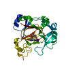
Mass: 146.098 Da / Num. of mol.: 1
Source method: isolated from a genetically manipulated source
Formula: C5H6O5
#3: Chemical ChemComp-NMM / (2S)-2-amino-5-[(N-methylcarbamimidoyl)amino]pentanoic acid / L-NMMA / Methylarginine


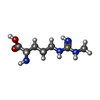
Type: L-peptide linking / Mass: 188.228 Da / Num. of mol.: 1 / Source method: obtained synthetically / Formula: C7H16N4O2 / Comment: inhibitor*YM
#4: Chemical ChemComp-ZN / ZINC ION


Mass: 65.409 Da / Num. of mol.: 1 / Source method: obtained synthetically / Formula: Zn
#5: Water ChemComp-HOH / water / Water


Mass: 18.015 Da / Num. of mol.: 90 / Source method: isolated from a natural source / Formula: H2O

-
Experimental details

-
Experiment

ExperimentMethod: X-RAY DIFFRACTION / Number of used crystals: 1

-
Sample preparation

CrystalDensity Matthews: 2.3 Å3/Da / Density % sol: 46.49 %
Crystal growTemperature: 281 K / Method: evaporation / Details: PEG3350 HEPEPS-Na / PH range: 7

-
Data collection

DiffractionMean temperature: 100 K
Diffraction sourceSource: ROTATING ANODE / Type: OTHER / Wavelength: 1.5418 Å
DetectorType: RIGAKU RAXIS IV++ / Detector: IMAGE PLATE / Date: Sep 3, 2015
RadiationProtocol: SINGLE WAVELENGTH / Monochromatic (M) / Laue (L): M / Scattering type: x-ray
Radiation wavelengthWavelength: 1.5418 Å / Relative weight: 1
ReflectionResolution: 2.4→38.975 Å / Num. obs: 7775 / % possible obs: 77.24 % / Redundancy: 6.7 % / Net I/σ(I): 2

-
Processing

Software
NameVersionClassification
PHENIX1.9_1692refinement
HKL-3000data reduction
HKL-3000data scaling
MOLREPphasing
RefinementMethod to determine structure: MOLECULAR REPLACEMENT
Starting model: 4QU1
Resolution: 2.42→38.975 Å / SU ML: 0.26 / Cross valid method: FREE R-VALUE / σ(F): 1.35 / Phase error: 24.07 / Stereochemistry target values: ML
RfactorNum. reflection% reflection
Rfree0.2458 372 4.78 %
Rwork0.1898 --
obs0.1925 7775 77.24 %
Solvent computationShrinkage radii: 0.9 Å / VDW probe radii: 1.11 Å / Solvent model: FLAT BULK SOLVENT MODEL
Refinement stepCycle: LAST / Resolution: 2.42→38.975 Å
ProteinNucleic acidLigandSolventTotal
Num. atoms1921 0 24 90 2035
Refine LS restraints
Refine-IDTypeDev idealNumber
X-RAY DIFFRACTIONf_bond_d0.0042012
X-RAY DIFFRACTIONf_angle_d0.9252744
X-RAY DIFFRACTIONf_dihedral_angle_d16.546740
X-RAY DIFFRACTIONf_chiral_restr0.038288
X-RAY DIFFRACTIONf_plane_restr0.004358
LS refinement shell
Resolution (Å)Rfactor RfreeNum. reflection RfreeRfactor RworkNum. reflection RworkRefine-ID% reflection obs (%)
2.4202-2.77040.31011360.23992529X-RAY DIFFRACTION81
2.7704-3.490.2281160.19442571X-RAY DIFFRACTION81
3.49-38.980.22671200.16412303X-RAY DIFFRACTION70

+
About Yorodumi

-
News

-
Feb 9, 2022. New format data for meta-information of EMDB entries

New format data for meta-information of EMDB entries

  • Version 3 of the EMDB header file is now the official format.
  • The previous official version 1.9 will be removed from the archive.

Related info.:EMDB header

External links:wwPDB to switch to version 3 of the EMDB data model

-
Aug 12, 2020. Covid-19 info

Covid-19 info

URL: https://pdbj.org/emnavi/covid19.php

New page: Covid-19 featured information page in EM Navigator.

Related info.:Covid-19 info / Mar 5, 2020. Novel coronavirus structure data

+
Mar 5, 2020. Novel coronavirus structure data

Novel coronavirus structure data

Related info.:Yorodumi Speices / Aug 12, 2020. Covid-19 info

External links:COVID-19 featured content - PDBj / Molecule of the Month (242):Coronavirus Proteases

+
Jan 31, 2019. EMDB accession codes are about to change! (news from PDBe EMDB page)

EMDB accession codes are about to change! (news from PDBe EMDB page)

  • The allocation of 4 digits for EMDB accession codes will soon come to an end. Whilst these codes will remain in use, new EMDB accession codes will include an additional digit and will expand incrementally as the available range of codes is exhausted. The current 4-digit format prefixed with “EMD-” (i.e. EMD-XXXX) will advance to a 5-digit format (i.e. EMD-XXXXX), and so on. It is currently estimated that the 4-digit codes will be depleted around Spring 2019, at which point the 5-digit format will come into force.
  • The EM Navigator/Yorodumi systems omit the EMD- prefix.

Related info.:Q: What is EMD? / ID/Accession-code notation in Yorodumi/EM Navigator

External links:EMDB Accession Codes are Changing Soon! / Contact to PDBj

+
Jul 12, 2017. Major update of PDB

Major update of PDB

  • wwPDB released updated PDB data conforming to the new PDBx/mmCIF dictionary.
  • This is a major update changing the version number from 4 to 5, and with Remediation, in which all the entries are updated.
  • In this update, many items about electron microscopy experimental information are reorganized (e.g. em_software).
  • Now, EM Navigator and Yorodumi are based on the updated data.

External links:wwPDB Remediation / Enriched Model Files Conforming to OneDep Data Standards Now Available in the PDB FTP Archive

-
Yorodumi

Thousand views of thousand structures

  • Yorodumi is a browser for structure data from EMDB, PDB, SASBDB, etc.
  • This page is also the successor to EM Navigator detail page, and also detail information page/front-end page for Omokage search.
  • The word "yorodu" (or yorozu) is an old Japanese word meaning "ten thousand". "mi" (miru) is to see.

Related info.:EMDB / PDB / SASBDB / Comparison of 3 databanks / Yorodumi Search / Aug 31, 2016. New EM Navigator & Yorodumi / Yorodumi Papers / Jmol/JSmol / Function and homology information / Changes in new EM Navigator and Yorodumi

Read more