 Movie
Movie Controller

[English] 日本語
 Yorodumi
- PDB-7dvv: Heme sensor protein PefR from Streptococcus agalactiae bound to o... -
+ Open data

- Basic information
| Entry | Database: PDB / ID: 7dvv | ||||||||||||
|---|---|---|---|---|---|---|---|---|---|---|---|---|---|
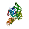
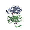
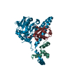
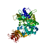
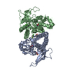
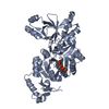
| Title | Heme sensor protein PefR from Streptococcus agalactiae bound to operator DNA (28-mer) | ||||||||||||
 Components Components |
| ||||||||||||
 Keywords Keywords | TRANSCRIPTION / heme-binding / helix-turn-helix | ||||||||||||
| Function / homology |  Function and homology information Function and homology informationresponse to stress / DNA-binding transcription factor activity / DNA binding / metal ion binding Similarity search - Function | ||||||||||||
| Biological species | Streptococcus agalactiae serotype III Streptococcus agalactiae NEM316 (bacteria) Streptococcus agalactiae NEM316 (bacteria) | ||||||||||||
| Method |  X-RAY DIFFRACTION / SYNCHROTRON / MOLECULAR REPLACEMENT / molecular replacement / Resolution: 2.49 Å X-RAY DIFFRACTION / SYNCHROTRON / MOLECULAR REPLACEMENT / molecular replacement / Resolution: 2.49 Å | ||||||||||||
| Model details | heme sensor protein | ||||||||||||
 Authors Authors | Nishinaga, M. / Nagai, S. / Nishitani, Y. / Sugimoto, H. / Shiro, Y. / Sawai, H. | ||||||||||||
| Funding support |  Japan, 3items Japan, 3items
| ||||||||||||
 Citation Citation | Journal: Commun Biol / Year: 2021 Title: Heme controls the structural rearrangement of its sensor protein mediating the hemolytic bacterial survival. Authors: Nishinaga, M. / Sugimoto, H. / Nishitani, Y. / Nagai, S. / Nagatoishi, S. / Muraki, N. / Tosha, T. / Tsumoto, K. / Aono, S. / Shiro, Y. / Sawai, H. | ||||||||||||
| History |
|
- Structure visualization
| Structure viewer | Molecule: MolmilJmol/JSmol |
|---|
- Downloads & links
-Download
| PDBx/mmCIF format | 7dvv.cif.gz | 192.4 KB | Display | PDBx/mmCIF format |
|---|---|---|---|---|
| PDB format | pdb7dvv.ent.gz | 148.2 KB | Display | PDB format |
| PDBx/mmJSON format | 7dvv.json.gz | Tree view | PDBx/mmJSON format | |
| Others |  Other downloads Other downloads |
-Validation report
| Arichive directory | https://data.pdbj.org/pub/pdb/validation_reports/dv/7dvvftp://data.pdbj.org/pub/pdb/validation_reports/dv/7dvv | HTTPS FTP |
|---|
-Related structure data
| Related structure data | 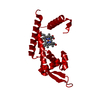 7dvrC  7dvsSC  7dvtC 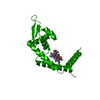 7dvuC  4llnS C: citing same article ( S: Starting model for refinement |
|---|---|
| Similar structure data |


-Links
 PDBj
PDBj


- Assembly
| Deposited unit | 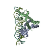
| ||||||||
|---|---|---|---|---|---|---|---|---|---|
| 1 |
| ||||||||
| Unit cell |
|
-Components
| #1: Protein | Mass: 17817.852 Da / Num. of mol.: 2 / Fragment: heme sensor protein Source method: isolated from a genetically manipulated source Source: (gene. exp.) Streptococcus agalactiae serotype III (strain NEM316) (bacteria)Strain: NEM316 / Gene: gbs1402 / Plasmid: pET-22b / Production host: #2: DNA chain | | Mass: 8579.605 Da / Num. of mol.: 1 / Source method: obtained synthetically / Source: (synth.) Streptococcus agalactiae NEM316 (bacteria)#3: DNA chain | | Mass: 8623.598 Da / Num. of mol.: 1 / Source method: obtained synthetically / Source: (synth.) Streptococcus agalactiae NEM316 (bacteria)#4: Water | ChemComp-HOH / |  Mass: 18.015 Da / Num. of mol.: 9 / Source method: isolated from a natural source / Formula: H2O Mass: 18.015 Da / Num. of mol.: 9 / Source method: isolated from a natural source / Formula: H2O |
|---|
-Experimental details
-Experiment
| Experiment | Method: X-RAY DIFFRACTION / Number of used crystals: 1 |
|---|
- Sample preparation
| Crystal | Density Matthews: 3.59 Å3/Da / Density % sol: 65.76 % |
|---|---|
| Crystal grow | Temperature: 293 K / Method: vapor diffusion, sitting drop / pH: 6.5 Details: 25%(w/v) PEGMME 2000, 0.2 M lithium sulfate, 0.1 M MES |
-Data collection
| Diffraction | Mean temperature: 100 K / Serial crystal experiment: N | ||||||||||||||||||||||||||||||||||||||||||||||||||||||||||||||||||||||||||||||||||||||||||||||||||||
|---|---|---|---|---|---|---|---|---|---|---|---|---|---|---|---|---|---|---|---|---|---|---|---|---|---|---|---|---|---|---|---|---|---|---|---|---|---|---|---|---|---|---|---|---|---|---|---|---|---|---|---|---|---|---|---|---|---|---|---|---|---|---|---|---|---|---|---|---|---|---|---|---|---|---|---|---|---|---|---|---|---|---|---|---|---|---|---|---|---|---|---|---|---|---|---|---|---|---|---|---|---|
| Diffraction source | Source: SYNCHROTRON / Site: SPring-8 / Beamline: BL41XU / Wavelength: 1 Å | ||||||||||||||||||||||||||||||||||||||||||||||||||||||||||||||||||||||||||||||||||||||||||||||||||||
| Detector | Type: DECTRIS EIGER X 16M / Detector: PIXEL / Date: Jan 22, 2018 / Details: mirrors | ||||||||||||||||||||||||||||||||||||||||||||||||||||||||||||||||||||||||||||||||||||||||||||||||||||
| Radiation | Monochromator: Si(111) / Protocol: SINGLE WAVELENGTH / Monochromatic (M) / Laue (L): M / Scattering type: x-ray | ||||||||||||||||||||||||||||||||||||||||||||||||||||||||||||||||||||||||||||||||||||||||||||||||||||
| Radiation wavelength | Wavelength: 1 Å / Relative weight: 1 | ||||||||||||||||||||||||||||||||||||||||||||||||||||||||||||||||||||||||||||||||||||||||||||||||||||
| Reflection | Resolution: 2.49→47.06 Å / Num. obs: 26423 / % possible obs: 99.9 % / Redundancy: 6.992 % / Biso Wilson estimate: 75.503 Å2 / CC1/2: 0.999 / Rmerge(I) obs: 0.083 / Rrim(I) all: 0.089 / Χ2: 1.321 / Net I/σ(I): 14.63 / Num. measured all: 357952 | ||||||||||||||||||||||||||||||||||||||||||||||||||||||||||||||||||||||||||||||||||||||||||||||||||||
| Reflection shell | Diffraction-ID: 1
|
-Phasing
| Phasing | Method: molecular replacement | |||||||||
|---|---|---|---|---|---|---|---|---|---|---|
| Phasing MR | Model details: Phaser MODE: MR_AUTO
|
- Processing
| Software |
| |||||||||||||||||||||||||||||||||||||||||||||||||||||||||||||||||||||||||||||||||||||||||||||||||||||||||||||||||||||||||||||
|---|---|---|---|---|---|---|---|---|---|---|---|---|---|---|---|---|---|---|---|---|---|---|---|---|---|---|---|---|---|---|---|---|---|---|---|---|---|---|---|---|---|---|---|---|---|---|---|---|---|---|---|---|---|---|---|---|---|---|---|---|---|---|---|---|---|---|---|---|---|---|---|---|---|---|---|---|---|---|---|---|---|---|---|---|---|---|---|---|---|---|---|---|---|---|---|---|---|---|---|---|---|---|---|---|---|---|---|---|---|---|---|---|---|---|---|---|---|---|---|---|---|---|---|---|---|---|
| Refinement | Method to determine structure: MOLECULAR REPLACEMENT Starting model: 7DVS, 4LLN Resolution: 2.49→47.06 Å / Cor.coef. Fo:Fc: 0.952 / Cor.coef. Fo:Fc free: 0.947 / SU B: 27.848 / SU ML: 0.256 / SU R Cruickshank DPI: 0.302 / Cross valid method: THROUGHOUT / σ(F): 0 / ESU R: 0.302 / ESU R Free: 0.231 Details: HYDROGENS HAVE BEEN USED IF PRESENT IN THE INPUT U VALUES : WITH TLS ADDED
| |||||||||||||||||||||||||||||||||||||||||||||||||||||||||||||||||||||||||||||||||||||||||||||||||||||||||||||||||||||||||||||
| Solvent computation | Ion probe radii: 0.8 Å / Shrinkage radii: 0.8 Å / VDW probe radii: 1.2 Å | |||||||||||||||||||||||||||||||||||||||||||||||||||||||||||||||||||||||||||||||||||||||||||||||||||||||||||||||||||||||||||||
| Displacement parameters | Biso max: 229.69 Å2 / Biso mean: 86.059 Å2 / Biso min: 32.13 Å2
| |||||||||||||||||||||||||||||||||||||||||||||||||||||||||||||||||||||||||||||||||||||||||||||||||||||||||||||||||||||||||||||
| Refinement step | Cycle: final / Resolution: 2.49→47.06 Å
| |||||||||||||||||||||||||||||||||||||||||||||||||||||||||||||||||||||||||||||||||||||||||||||||||||||||||||||||||||||||||||||
| Refine LS restraints |
| |||||||||||||||||||||||||||||||||||||||||||||||||||||||||||||||||||||||||||||||||||||||||||||||||||||||||||||||||||||||||||||
| LS refinement shell | Resolution: 2.495→2.559 Å / Rfactor Rfree error: 0 / Total num. of bins used: 20
| |||||||||||||||||||||||||||||||||||||||||||||||||||||||||||||||||||||||||||||||||||||||||||||||||||||||||||||||||||||||||||||
| Refinement TLS params. | Method: refined / Refine-ID: X-RAY DIFFRACTION
| |||||||||||||||||||||||||||||||||||||||||||||||||||||||||||||||||||||||||||||||||||||||||||||||||||||||||||||||||||||||||||||
| Refinement TLS group |
|
