 Movie
Movie Controller

[English] 日本語
 Yorodumi
- PDB-7doa: Crystal structure of Catabolite repressor acivator from E. coli i... -
+ Open data

- Basic information
| Entry | Database: PDB / ID: 7doa | ||||||
|---|---|---|---|---|---|---|---|
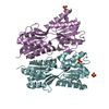
| Title | Crystal structure of Catabolite repressor acivator from E. coli in complex with HEPES | ||||||
 Components Components | Catabolite repressor/activator | ||||||
 Keywords Keywords | GENE REGULATION / Cra / HEPES / DNA BINDING PROTEIN | ||||||
| Function / homology |  Function and homology information Function and homology informationresponse to fructose / transcription cis-regulatory region binding / DNA-binding transcription factor activity Similarity search - Function | ||||||
| Biological species |  | ||||||
| Method |  X-RAY DIFFRACTION / SYNCHROTRON / MOLECULAR REPLACEMENT / Resolution: 2.7 Å X-RAY DIFFRACTION / SYNCHROTRON / MOLECULAR REPLACEMENT / Resolution: 2.7 Å | ||||||
 Authors Authors | Neetu, N. / Katiki, M. / Kumar, P. | ||||||
 Citation Citation | Journal: To Be Published Title: Crystal structure of Catabolite repressor acivator from E. coli in complex with HEPES Authors: Neetu, N. / Katiki, M. / Kumar, P. | ||||||
| History |
|
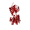
- Structure visualization
| Structure viewer | Molecule: MolmilJmol/JSmol |
|---|
- Downloads & links
-Download
| PDBx/mmCIF format | 7doa.cif.gz | 244.3 KB | Display | PDBx/mmCIF format |
|---|---|---|---|---|
| PDB format | pdb7doa.ent.gz | 197.3 KB | Display | PDB format |
| PDBx/mmJSON format | 7doa.json.gz | Tree view | PDBx/mmJSON format | |
| Others |  Other downloads Other downloads |
-Validation report
| Summary document | 7doa_validation.pdf.gz | 1.1 MB | Display | wwPDB validaton report |
|---|---|---|---|---|
| Full document | 7doa_full_validation.pdf.gz | 1.1 MB | Display | |
| Data in XML | 7doa_validation.xml.gz | 24.9 KB | Display | |
| Data in CIF | 7doa_validation.cif.gz | 33.2 KB | Display | |
| Arichive directory | https://data.pdbj.org/pub/pdb/validation_reports/do/7doaftp://data.pdbj.org/pub/pdb/validation_reports/do/7doa | HTTPS FTP |
-Related structure data
| Related structure data | 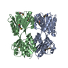 2iksS S: Starting model for refinement |
|---|---|
| Similar structure data |


-Links
 PDBj
PDBj


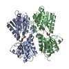
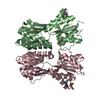
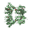
- Assembly
| Deposited unit | 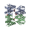
| |||||||||||||||||||||||||||||||||
|---|---|---|---|---|---|---|---|---|---|---|---|---|---|---|---|---|---|---|---|---|---|---|---|---|---|---|---|---|---|---|---|---|---|---|
| 1 |
| |||||||||||||||||||||||||||||||||
| Unit cell |
| |||||||||||||||||||||||||||||||||
| Components on special symmetry positions |
| |||||||||||||||||||||||||||||||||
| Noncrystallographic symmetry (NCS) | NCS domain:
NCS domain segments: Ens-ID: 1 / Beg auth comp-ID: ARG / Beg label comp-ID: ARG / End auth comp-ID: ARG / End label comp-ID: ARG / Auth seq-ID: 60 - 333 / Label seq-ID: 83 - 356
NCS oper:
|
-Components
| #1: Protein | Mass: 40811.289 Da / Num. of mol.: 2 Source method: isolated from a genetically manipulated source Source: (gene. exp.) Strain: 536 / UPEC / Gene: ECP_0082 / Production host: #2: Chemical |   Mass: 238.305 Da / Num. of mol.: 3 / Source method: obtained synthetically / Formula: C8H18N2O4S / Feature type: SUBJECT OF INVESTIGATION / Comment: pH buffer*YM Mass: 238.305 Da / Num. of mol.: 3 / Source method: obtained synthetically / Formula: C8H18N2O4S / Feature type: SUBJECT OF INVESTIGATION / Comment: pH buffer*YM#3: Chemical | ChemComp-GOL / |   Mass: 92.094 Da / Num. of mol.: 1 / Source method: obtained synthetically / Formula: C3H8O3 Mass: 92.094 Da / Num. of mol.: 1 / Source method: obtained synthetically / Formula: C3H8O3#4: Water | ChemComp-HOH / |  Mass: 18.015 Da / Num. of mol.: 63 / Source method: isolated from a natural source / Formula: H2O Mass: 18.015 Da / Num. of mol.: 63 / Source method: isolated from a natural source / Formula: H2OHas ligand of interest | Y | |
|---|
-Experimental details
-Experiment
| Experiment | Method: X-RAY DIFFRACTION / Number of used crystals: 1 |
|---|
- Sample preparation
| Crystal | Density Matthews: 1.79 Å3/Da / Density % sol: 31.42 % |
|---|---|
| Crystal grow | Temperature: 293 K / Method: vapor diffusion / Details: HEPES, MgCl2, PEG 4000 |
-Data collection
| Diffraction | Mean temperature: 100 K / Serial crystal experiment: N |
|---|---|
| Diffraction source | Source: SYNCHROTRON / Site: ESRF  / Beamline: ID23-2 / Wavelength: 0.873 Å / Beamline: ID23-2 / Wavelength: 0.873 Å |
| Detector | Type: DECTRIS PILATUS 300K / Detector: PIXEL / Date: Sep 1, 2017 |
| Radiation | Protocol: SINGLE WAVELENGTH / Monochromatic (M) / Laue (L): M / Scattering type: x-ray |
| Radiation wavelength | Wavelength: 0.873 Å / Relative weight: 1 |
| Reflection | Resolution: 2.7→40.91 Å / Num. obs: 16445 / % possible obs: 97.78 % / Redundancy: 0.08726 % / CC1/2: 0.998 / Rmerge(I) obs: 0.074 / Net I/σ(I): 10.6 |
| Reflection shell | Resolution: 2.7→2.797 Å / Num. unique obs: 3740 / CC1/2: 0.9 |
- Processing
| Software |
| |||||||||||||||||||||||||||||||||||||||||||||||||||||||||||||||||||||||||||
|---|---|---|---|---|---|---|---|---|---|---|---|---|---|---|---|---|---|---|---|---|---|---|---|---|---|---|---|---|---|---|---|---|---|---|---|---|---|---|---|---|---|---|---|---|---|---|---|---|---|---|---|---|---|---|---|---|---|---|---|---|---|---|---|---|---|---|---|---|---|---|---|---|---|---|---|---|
| Refinement | Method to determine structure: MOLECULAR REPLACEMENT Starting model: 2IKS Resolution: 2.7→40.91 Å / Cor.coef. Fo:Fc: 0.952 / Cor.coef. Fo:Fc free: 0.923 / SU B: 49.063 / SU ML: 0.446 / Cross valid method: THROUGHOUT / σ(F): 0 / ESU R Free: 0.425 / Stereochemistry target values: MAXIMUM LIKELIHOOD / Details: U VALUES : WITH TLS ADDED
| |||||||||||||||||||||||||||||||||||||||||||||||||||||||||||||||||||||||||||
| Solvent computation | Ion probe radii: 0.8 Å / Shrinkage radii: 0.8 Å / VDW probe radii: 1.2 Å / Solvent model: MASK | |||||||||||||||||||||||||||||||||||||||||||||||||||||||||||||||||||||||||||
| Displacement parameters | Biso max: 147.87 Å2 / Biso mean: 74.035 Å2 / Biso min: 41.27 Å2
| |||||||||||||||||||||||||||||||||||||||||||||||||||||||||||||||||||||||||||
| Refinement step | Cycle: final / Resolution: 2.7→40.91 Å
| |||||||||||||||||||||||||||||||||||||||||||||||||||||||||||||||||||||||||||
| Refine LS restraints |
| |||||||||||||||||||||||||||||||||||||||||||||||||||||||||||||||||||||||||||
| Refine LS restraints NCS | Ens-ID: 1 / Number: 16498 / Refine-ID: X-RAY DIFFRACTION / Type: interatomic distance / Rms dev position: 0.17 Å / Weight position: 0.05
| |||||||||||||||||||||||||||||||||||||||||||||||||||||||||||||||||||||||||||
| LS refinement shell | Resolution: 2.7→2.77 Å / Rfactor Rfree error: 0 / Total num. of bins used: 20
| |||||||||||||||||||||||||||||||||||||||||||||||||||||||||||||||||||||||||||
| Refinement TLS params. | Method: refined / Refine-ID: X-RAY DIFFRACTION
| |||||||||||||||||||||||||||||||||||||||||||||||||||||||||||||||||||||||||||
| Refinement TLS group |
|
