 Movie
Movie Controller

[English] 日本語
 Yorodumi
- PDB-7dma: Crystal structure of FliM middle domain (46-231) with R49P substi... -
+ Open data

- Basic information
| Entry | Database: PDB / ID: 7dma | ||||||||||||||||||
|---|---|---|---|---|---|---|---|---|---|---|---|---|---|---|---|---|---|---|---|
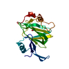
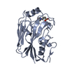
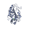
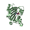
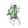
| Title | Crystal structure of FliM middle domain (46-231) with R49P substitution from Vibro alginolyticus | ||||||||||||||||||
 Components Components | (Flagellar motor switch protein FliM) x 2 | ||||||||||||||||||
 Keywords Keywords | MOTOR PROTEIN / Flagellar motor protein | ||||||||||||||||||
| Function / homology |  Function and homology information Function and homology informationbacterial-type flagellum basal body / cytoskeletal motor activity / bacterial-type flagellum-dependent cell motility / chemotaxis / plasma membrane Similarity search - Function | ||||||||||||||||||
| Biological species |  Vibrio alginolyticus (bacteria) Vibrio alginolyticus (bacteria) | ||||||||||||||||||
| Method |  X-RAY DIFFRACTION / SYNCHROTRON / MOLECULAR REPLACEMENT / Resolution: 1.44 Å X-RAY DIFFRACTION / SYNCHROTRON / MOLECULAR REPLACEMENT / Resolution: 1.44 Å | ||||||||||||||||||
 Authors Authors | Takekawa, N. / Homma, M. / Imada, K. | ||||||||||||||||||
| Funding support |  Japan, 5items Japan, 5items
| ||||||||||||||||||
 Citation Citation | Journal: J.Biochem. / Year: 2021 Title: A slight bending of an alpha-helix in FliM creates a counterclockwise-locked structure of the flagellar motor in Vibrio. Authors: Takekawa, N. / Nishikino, T. / Yamashita, T. / Hori, K. / Onoue, Y. / Ihara, K. / Kojima, S. / Homma, M. / Imada, K. | ||||||||||||||||||
| History |
|
- Structure visualization
| Structure viewer | Molecule: MolmilJmol/JSmol |
|---|
- Downloads & links
-Download
| PDBx/mmCIF format | 7dma.cif.gz | 63.4 KB | Display | PDBx/mmCIF format |
|---|---|---|---|---|
| PDB format | pdb7dma.ent.gz | 41.1 KB | Display | PDB format |
| PDBx/mmJSON format | 7dma.json.gz | Tree view | PDBx/mmJSON format | |
| Others |  Other downloads Other downloads |
-Validation report
| Summary document | 7dma_validation.pdf.gz | 437.7 KB | Display | wwPDB validaton report |
|---|---|---|---|---|
| Full document | 7dma_full_validation.pdf.gz | 440.3 KB | Display | |
| Data in XML | 7dma_validation.xml.gz | 10.9 KB | Display | |
| Data in CIF | 7dma_validation.cif.gz | 15.2 KB | Display | |
| Arichive directory | https://data.pdbj.org/pub/pdb/validation_reports/dm/7dmaftp://data.pdbj.org/pub/pdb/validation_reports/dm/7dma | HTTPS FTP |
-Related structure data
| Related structure data | 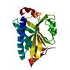 7dm9C 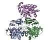 5x0zS C: citing same article ( S: Starting model for refinement |
|---|---|
| Similar structure data |


-Links
 PDBj
PDBj- Assembly
| Deposited unit | 
| ||||||||||||
|---|---|---|---|---|---|---|---|---|---|---|---|---|---|
| 1 |
| ||||||||||||
| Unit cell |
|
-Components
| #1: Protein | Mass: 11921.076 Da / Num. of mol.: 1 / Fragment: UNP residues 42-140 / Mutation: R49P Source method: isolated from a genetically manipulated source Source: (gene. exp.) Vibrio alginolyticus (bacteria) / Gene: fliM / Plasmid: pET15b / Production host: |
|---|---|
| #2: Protein | Mass: 10310.676 Da / Num. of mol.: 1 / Fragment: UNP residues 141-231 Source method: isolated from a genetically manipulated source Source: (gene. exp.) Vibrio alginolyticus (bacteria) / Gene: fliM / Plasmid: pET15b / Production host: |
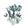
| #3: Water | ChemComp-HOH /  Mass: 18.015 Da / Num. of mol.: 170 / Source method: isolated from a natural source / Formula: H2O Mass: 18.015 Da / Num. of mol.: 170 / Source method: isolated from a natural source / Formula: H2O |
| Sequence details | The single protein was digested at the position between A138 and E141. The distance between A138 ...The single protein was digested at the position between A138 and E141. The distance between A138 and E141 is 30.61 Angstrom. Therefore the polymer has been split in to two, chain A and chain B for curation. |
-Experimental details
-Experiment
| Experiment | Method: X-RAY DIFFRACTION / Number of used crystals: 1 |
|---|
- Sample preparation
| Crystal | Density Matthews: 1.67 Å3/Da / Density % sol: 22.5 % |
|---|---|
| Crystal grow | Temperature: 293 K / Method: vapor diffusion, sitting drop / pH: 7.5 / Details: 30 % (w/v) PEG-400, 0.1 M HEPES-NaOH, 0.2 M NaCl |
-Data collection
| Diffraction | Mean temperature: 100 K / Serial crystal experiment: N |
|---|---|
| Diffraction source | Source: SYNCHROTRON / Site: SPring-8 / Beamline: BL41XU / Wavelength: 1 Å |
| Detector | Type: DECTRIS EIGER X 16M / Detector: PIXEL / Date: Nov 29, 2019 |
| Radiation | Monochromator: Double-crystal monochromator / Protocol: SINGLE WAVELENGTH / Monochromatic (M) / Laue (L): M / Scattering type: x-ray |
| Radiation wavelength | Wavelength: 1 Å / Relative weight: 1 |
| Reflection | Resolution: 1.44→75.87 Å / Num. obs: 26243 / % possible obs: 100 % / Redundancy: 6.1 % / Biso Wilson estimate: 16.14 Å2 / CC1/2: 0.996 / Rmerge(I) obs: 0.078 / Rpim(I) all: 0.049 / Rrim(I) all: 0.093 / Net I/σ(I): 10.5 |
| Reflection shell | Resolution: 1.44→1.47 Å / Redundancy: 5.6 % / Rmerge(I) obs: 0.481 / Mean I/σ(I) obs: 2.6 / Num. unique obs: 1332 / CC1/2: 0.881 / Rpim(I) all: 0.307 / Rrim(I) all: 0.574 / % possible all: 100 |
- Processing
| Software |
| |||||||||||||||||||||||||||||||||||||||||||||||||||||||||||||||||||||||||||||||||||||||||||||||||||||||||
|---|---|---|---|---|---|---|---|---|---|---|---|---|---|---|---|---|---|---|---|---|---|---|---|---|---|---|---|---|---|---|---|---|---|---|---|---|---|---|---|---|---|---|---|---|---|---|---|---|---|---|---|---|---|---|---|---|---|---|---|---|---|---|---|---|---|---|---|---|---|---|---|---|---|---|---|---|---|---|---|---|---|---|---|---|---|---|---|---|---|---|---|---|---|---|---|---|---|---|---|---|---|---|---|---|---|---|
| Refinement | Method to determine structure: MOLECULAR REPLACEMENT Starting model: 5X0Z Resolution: 1.44→48.46 Å / SU ML: 0.1511 / Cross valid method: FREE R-VALUE / σ(F): 1.36 / Phase error: 19.6042 Stereochemistry target values: GeoStd + Monomer Library + CDL v1.2
| |||||||||||||||||||||||||||||||||||||||||||||||||||||||||||||||||||||||||||||||||||||||||||||||||||||||||
| Solvent computation | Shrinkage radii: 0.9 Å / VDW probe radii: 1.11 Å / Solvent model: FLAT BULK SOLVENT MODEL | |||||||||||||||||||||||||||||||||||||||||||||||||||||||||||||||||||||||||||||||||||||||||||||||||||||||||
| Displacement parameters | Biso mean: 21.34 Å2 | |||||||||||||||||||||||||||||||||||||||||||||||||||||||||||||||||||||||||||||||||||||||||||||||||||||||||
| Refinement step | Cycle: LAST / Resolution: 1.44→48.46 Å
| |||||||||||||||||||||||||||||||||||||||||||||||||||||||||||||||||||||||||||||||||||||||||||||||||||||||||
| Refine LS restraints |
| |||||||||||||||||||||||||||||||||||||||||||||||||||||||||||||||||||||||||||||||||||||||||||||||||||||||||
| LS refinement shell |
|
