 Movie
Movie Controller

[English] 日本語
 Yorodumi
- PDB-7dm9: Crystal structure of FliM middle domain (51-229) from Vibro algin... -
+ Open data

- Basic information
| Entry | Database: PDB / ID: 7dm9 | ||||||||||||||||||
|---|---|---|---|---|---|---|---|---|---|---|---|---|---|---|---|---|---|---|---|
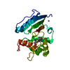
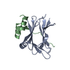
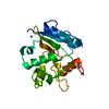
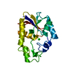
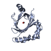
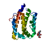
| Title | Crystal structure of FliM middle domain (51-229) from Vibro alginolyticus | ||||||||||||||||||
 Components Components | Flagellar motor switch protein FliM | ||||||||||||||||||
 Keywords Keywords | MOTOR PROTEIN / Flagellar motor protein | ||||||||||||||||||
| Function / homology |  Function and homology information Function and homology informationbacterial-type flagellum basal body / cytoskeletal motor activity / bacterial-type flagellum-dependent cell motility / chemotaxis / plasma membrane Similarity search - Function | ||||||||||||||||||
| Biological species |  Vibrio alginolyticus (bacteria) Vibrio alginolyticus (bacteria) | ||||||||||||||||||
| Method |  X-RAY DIFFRACTION / SYNCHROTRON / MOLECULAR REPLACEMENT / Resolution: 1.71 Å X-RAY DIFFRACTION / SYNCHROTRON / MOLECULAR REPLACEMENT / Resolution: 1.71 Å | ||||||||||||||||||
 Authors Authors | Takekawa, N. / Homma, M. / Imada, K. | ||||||||||||||||||
| Funding support |  Japan, 5items Japan, 5items
| ||||||||||||||||||
 Citation Citation | Journal: J.Biochem. / Year: 2021 Title: A slight bending of an alpha-helix in FliM creates a counterclockwise-locked structure of the flagellar motor in Vibrio. Authors: Takekawa, N. / Nishikino, T. / Yamashita, T. / Hori, K. / Onoue, Y. / Ihara, K. / Kojima, S. / Homma, M. / Imada, K. | ||||||||||||||||||
| History |
|
- Structure visualization
| Structure viewer | Molecule: MolmilJmol/JSmol |
|---|
- Downloads & links
-Download
| PDBx/mmCIF format | 7dm9.cif.gz | 56.7 KB | Display | PDBx/mmCIF format |
|---|---|---|---|---|
| PDB format | pdb7dm9.ent.gz | 35.3 KB | Display | PDB format |
| PDBx/mmJSON format | 7dm9.json.gz | Tree view | PDBx/mmJSON format | |
| Others |  Other downloads Other downloads |
-Validation report
| Summary document | 7dm9_validation.pdf.gz | 423.7 KB | Display | wwPDB validaton report |
|---|---|---|---|---|
| Full document | 7dm9_full_validation.pdf.gz | 424.6 KB | Display | |
| Data in XML | 7dm9_validation.xml.gz | 9.3 KB | Display | |
| Data in CIF | 7dm9_validation.cif.gz | 12.6 KB | Display | |
| Arichive directory | https://data.pdbj.org/pub/pdb/validation_reports/dm/7dm9ftp://data.pdbj.org/pub/pdb/validation_reports/dm/7dm9 | HTTPS FTP |
-Related structure data
| Related structure data | 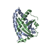 7dmaC 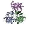 5x0zS S: Starting model for refinement C: citing same article ( |
|---|---|
| Similar structure data |


-Links
 PDBj
PDBj- Assembly
| Deposited unit | 
| ||||||||||||
|---|---|---|---|---|---|---|---|---|---|---|---|---|---|
| 1 |
| ||||||||||||
| Unit cell |
|
-Components
| #1: Protein | Mass: 22273.840 Da / Num. of mol.: 1 Source method: isolated from a genetically manipulated source Details: The N-terminal GSHM is a remnant of the His-tag / Source: (gene. exp.) Vibrio alginolyticus (bacteria)Gene: Vag1382_20640, VagVIO5_20640, VagYM19_20670, VagYM4_20660 Plasmid: pET15b / Production host: |
|---|---|
| #2: Water | ChemComp-HOH /  Mass: 18.015 Da / Num. of mol.: 98 / Source method: isolated from a natural source / Formula: H2O Mass: 18.015 Da / Num. of mol.: 98 / Source method: isolated from a natural source / Formula: H2O |
-Experimental details
-Experiment
| Experiment | Method: X-RAY DIFFRACTION / Number of used crystals: 1 |
|---|
- Sample preparation
| Crystal | Density Matthews: 1.53 Å3/Da / Density % sol: 19.92 % |
|---|---|
| Crystal grow | Temperature: 293 K / Method: vapor diffusion, sitting drop / pH: 9.5 / Details: 40 % (w/v) PEG-600, 0.1 M CHES-NaOH |
-Data collection
| Diffraction | Mean temperature: 100 K / Serial crystal experiment: N |
|---|---|
| Diffraction source | Source: SYNCHROTRON / Site: SPring-8 / Beamline: BL41XU / Wavelength: 1 Å |
| Detector | Type: DECTRIS EIGER X 16M / Detector: PIXEL / Date: Nov 29, 2019 |
| Radiation | Monochromator: Double-crystal monochromator / Protocol: SINGLE WAVELENGTH / Monochromatic (M) / Laue (L): M / Scattering type: x-ray |
| Radiation wavelength | Wavelength: 1 Å / Relative weight: 1 |
| Reflection | Resolution: 1.71→73.89 Å / Num. obs: 15496 / % possible obs: 100 % / Redundancy: 5.9 % / Biso Wilson estimate: 19.4 Å2 / CC1/2: 0.996 / Rmerge(I) obs: 0.066 / Rpim(I) all: 0.044 / Rrim(I) all: 0.079 / Net I/σ(I): 14.3 |
| Reflection shell | Resolution: 1.71→1.74 Å / Redundancy: 6 % / Rmerge(I) obs: 0.264 / Mean I/σ(I) obs: 5.7 / Num. unique obs: 824 / CC1/2: 0.959 / Rpim(I) all: 0.176 / Rrim(I) all: 0.319 / % possible all: 100 |
- Processing
| Software |
| ||||||||||||||||||||||||||||||||||||||||||
|---|---|---|---|---|---|---|---|---|---|---|---|---|---|---|---|---|---|---|---|---|---|---|---|---|---|---|---|---|---|---|---|---|---|---|---|---|---|---|---|---|---|---|---|
| Refinement | Method to determine structure: MOLECULAR REPLACEMENT Starting model: 5X0Z Resolution: 1.71→47.72 Å / SU ML: 0.1761 / Cross valid method: FREE R-VALUE / σ(F): 1.34 / Phase error: 21.7224 Stereochemistry target values: GeoStd + Monomer Library + CDL v1.2
| ||||||||||||||||||||||||||||||||||||||||||
| Solvent computation | Shrinkage radii: 0.9 Å / VDW probe radii: 1.11 Å / Solvent model: FLAT BULK SOLVENT MODEL | ||||||||||||||||||||||||||||||||||||||||||
| Displacement parameters | Biso mean: 23.33 Å2 | ||||||||||||||||||||||||||||||||||||||||||
| Refinement step | Cycle: LAST / Resolution: 1.71→47.72 Å
| ||||||||||||||||||||||||||||||||||||||||||
| Refine LS restraints |
| ||||||||||||||||||||||||||||||||||||||||||
| LS refinement shell |
|
