 Movie
Movie Controller


+ Open data

- Basic information
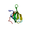
| Entry | Database: PDB / ID: 7da8 | ||||||
|---|---|---|---|---|---|---|---|
| Title | X-ray structure of a GB1:T2Q/D46K mutant | ||||||
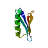
 Components Components | Immunoglobulin G-binding protein G | ||||||
 Keywords Keywords | PROTEIN BINDING / GB1 / T2Q mutant / D46K mutant / X-ray crystal structure / PLANT PROTEIN | ||||||
| Function / homology |  Function and homology information Function and homology information | ||||||
| Biological species |  Streptococcus sp. group G (bacteria) Streptococcus sp. group G (bacteria) | ||||||
| Method |  X-RAY DIFFRACTION / MOLECULAR REPLACEMENT / Resolution: 2.4 Å X-RAY DIFFRACTION / MOLECULAR REPLACEMENT / Resolution: 2.4 Å | ||||||
 Authors Authors | Manjula, R. / Ramaswamy, S. / Gosavi, S. | ||||||
 Citation Citation | Journal: To Be Published Title: X-ray structure of a GB1:T2Q/D46K mutant Authors: Manjula, R. / Ramaswamy, S. / Gosavi, S. | ||||||
| History |
|
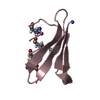
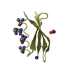
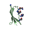
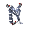
- Structure visualization
| Structure viewer | Molecule: MolmilJmol/JSmol |
|---|
- Downloads & links
-Download
| PDBx/mmCIF format | 7da8.cif.gz | 23.3 KB | Display | PDBx/mmCIF format |
|---|---|---|---|---|
| PDB format | pdb7da8.ent.gz | 13 KB | Display | PDB format |
| PDBx/mmJSON format | 7da8.json.gz | Tree view | PDBx/mmJSON format | |
| Others |  Other downloads Other downloads |
-Validation report
| Summary document | 7da8_validation.pdf.gz | 434.3 KB | Display | wwPDB validaton report |
|---|---|---|---|---|
| Full document | 7da8_full_validation.pdf.gz | 435.4 KB | Display | |
| Data in XML | 7da8_validation.xml.gz | 4.2 KB | Display | |
| Data in CIF | 7da8_validation.cif.gz | 4.9 KB | Display | |
| Arichive directory | https://data.pdbj.org/pub/pdb/validation_reports/da/7da8ftp://data.pdbj.org/pub/pdb/validation_reports/da/7da8 | HTTPS FTP |
-Related structure data
| Related structure data | 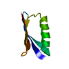 1pgbS S: Starting model for refinement |
|---|---|
| Similar structure data |


-Links
 PDBj
PDBj

- Assembly
| Deposited unit | 
| ||||||||
|---|---|---|---|---|---|---|---|---|---|
| 1 |
| ||||||||
| Unit cell |
|
-Components
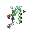
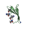
| #1: Antibody | Mass: 7071.782 Da / Num. of mol.: 1 / Mutation: T303Q, D347K Source method: isolated from a genetically manipulated source Source: (gene. exp.) Streptococcus sp. group G (bacteria) / Gene: spg / Production host: |
|---|---|
| #2: Water | ChemComp-HOH /  Mass: 18.015 Da / Num. of mol.: 7 / Source method: isolated from a natural source / Formula: H2O Mass: 18.015 Da / Num. of mol.: 7 / Source method: isolated from a natural source / Formula: H2O |
-Experimental details
-Experiment
| Experiment | Method: X-RAY DIFFRACTION / Number of used crystals: 1 |
|---|
- Sample preparation
| Crystal | Density Matthews: 1.66 Å3/Da / Density % sol: 26.03 % |
|---|---|
| Crystal grow | Temperature: 293 K / Method: vapor diffusion, sitting drop / pH: 6.5 Details: MES monohydrate pH 6.5 (buffer system 1), 40% v/v PEG 500* MME, 20 % w/v PEG 20000 (Precipitant mix1) (Morpheus Screen ID 25). |
-Data collection
| Diffraction | Mean temperature: 100 K / Serial crystal experiment: N | ||||||||||||||||||||||||||||||
|---|---|---|---|---|---|---|---|---|---|---|---|---|---|---|---|---|---|---|---|---|---|---|---|---|---|---|---|---|---|---|---|
| Diffraction source | Source: ROTATING ANODE / Type: RIGAKU R-AXIS / Wavelength: 1.54187 Å | ||||||||||||||||||||||||||||||
| Detector | Type: RIGAKU RAXIS IV++ / Detector: IMAGE PLATE / Date: Sep 6, 2019 | ||||||||||||||||||||||||||||||
| Radiation | Protocol: SINGLE WAVELENGTH / Monochromatic (M) / Laue (L): M / Scattering type: x-ray | ||||||||||||||||||||||||||||||
| Radiation wavelength | Wavelength: 1.54187 Å / Relative weight: 1 | ||||||||||||||||||||||||||||||
| Reflection | Resolution: 2.4→30.82 Å / Num. obs: 2003 / % possible obs: 98 % / Redundancy: 6.3 % / CC1/2: 0.973 / Rmerge(I) obs: 0.2 / Rpim(I) all: 0.093 / Rrim(I) all: 0.233 / Net I/σ(I): 5.9 | ||||||||||||||||||||||||||||||
| Reflection shell | Diffraction-ID: 1
|
- Processing
| Software |
| ||||||||||||||||||||||||
|---|---|---|---|---|---|---|---|---|---|---|---|---|---|---|---|---|---|---|---|---|---|---|---|---|---|
| Refinement | Method to determine structure: MOLECULAR REPLACEMENT Starting model: 1pgb Resolution: 2.4→30.819 Å / SU ML: 0.26 / Cross valid method: THROUGHOUT / σ(F): 1.38 / Phase error: 15.17 / Stereochemistry target values: ML
| ||||||||||||||||||||||||
| Solvent computation | Shrinkage radii: 0.9 Å / VDW probe radii: 1.11 Å / Solvent model: FLAT BULK SOLVENT MODEL | ||||||||||||||||||||||||
| Displacement parameters | Biso max: 76.7 Å2 / Biso mean: 33.7382 Å2 / Biso min: 12.19 Å2 | ||||||||||||||||||||||||
| Refinement step | Cycle: final / Resolution: 2.4→30.819 Å
| ||||||||||||||||||||||||
| LS refinement shell | Resolution: 2.406→30.81 Å / Rfactor Rfree error: 0
|
