 Movie
Movie Controller

[English] 日本語
 Yorodumi
- PDB-7d95: Crystal structure of acivicin-bound GATase subunit of Methanocald... -
+ Open data

- Basic information
| Entry | Database: PDB / ID: 7d95 | |||||||||
|---|---|---|---|---|---|---|---|---|---|---|
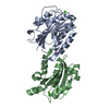
| Title | Crystal structure of acivicin-bound GATase subunit of Methanocaldococcus jannaschii GMP synthetase | |||||||||
 Components Components | GMP synthase [glutamine-hydrolyzing] subunit A | |||||||||
 Keywords Keywords | LIGASE / Succinimide / Deamidation / hyperthermostable glutamine amidotransferase / acivicin bound GATase | |||||||||
| Function / homology |  Function and homology information Function and homology informationGMP synthase (glutamine-hydrolyzing) activity / GMP synthase (glutamine-hydrolysing) / ATP binding Similarity search - Function | |||||||||
| Biological species |   Methanocaldococcus jannaschii DSM 2661 (archaea) Methanocaldococcus jannaschii DSM 2661 (archaea) | |||||||||
| Method |  X-RAY DIFFRACTION / MOLECULAR REPLACEMENT / Resolution: 1.67 Å X-RAY DIFFRACTION / MOLECULAR REPLACEMENT / Resolution: 1.67 Å | |||||||||
 Authors Authors | Bellur, A. / Dongre, A. / Kumar, S. / Balaram, H. | |||||||||
| Funding support |  India, 2items India, 2items
| |||||||||
 Citation Citation | Journal: Biophys.J. / Year: 2021 Title: Structural basis for the hyperthermostability of an archaeal enzyme induced by succinimide formation. Authors: Dongre, A.V. / Das, S. / Bellur, A. / Kumar, S. / Chandrashekarmath, A. / Karmakar, T. / Balaram, P. / Balasubramanian, S. / Balaram, H. | |||||||||
| History |
|
- Structure visualization
| Structure viewer | Molecule: MolmilJmol/JSmol |
|---|
- Downloads & links
-Download
| PDBx/mmCIF format | 7d95.cif.gz | 93.9 KB | Display | PDBx/mmCIF format |
|---|---|---|---|---|
| PDB format | pdb7d95.ent.gz | 69.7 KB | Display | PDB format |
| PDBx/mmJSON format | 7d95.json.gz | Tree view | PDBx/mmJSON format | |
| Others |  Other downloads Other downloads |
-Validation report
| Arichive directory | https://data.pdbj.org/pub/pdb/validation_reports/d9/7d95ftp://data.pdbj.org/pub/pdb/validation_reports/d9/7d95 | HTTPS FTP |
|---|
-Related structure data
| Related structure data |  7d40C  7d96C  7d97C  1wl8S S: Starting model for refinement C: citing same article ( |
|---|---|
| Similar structure data |


-Links
 PDBj
PDBj

- Assembly
| Deposited unit | 
| ||||||||
|---|---|---|---|---|---|---|---|---|---|
| 1 |
| ||||||||
| Unit cell |
|
-Components
| #1: Protein | Mass: 21177.363 Da / Num. of mol.: 2 Source method: isolated from a genetically manipulated source Source: (gene. exp.) Methanocaldococcus jannaschii DSM 2661 (archaea)Strain: DSM 2661 / Gene: guaAA, MJ1575 / Production host:  References: UniProt: Q58970, GMP synthase (glutamine-hydrolysing) #2: Water | ChemComp-HOH / |  Mass: 18.015 Da / Num. of mol.: 266 / Source method: isolated from a natural source / Formula: H2O Mass: 18.015 Da / Num. of mol.: 266 / Source method: isolated from a natural source / Formula: H2OHas ligand of interest | Y | |
|---|
-Experimental details
-Experiment
| Experiment | Method: X-RAY DIFFRACTION / Number of used crystals: 1 |
|---|
- Sample preparation
| Crystal | Density Matthews: 2.83 Å3/Da / Density % sol: 56.6 % Description: THE ENTRY CONTAINS FRIEDEL PAIRS IN I/F_PLUS/MINUS COLUMNS. |
|---|---|
| Crystal grow | Temperature: 277 K / Method: microbatch / pH: 4.6 / Details: 100 mM Sodium acetate, pH 4.6, 35 % PEG 6000 |
-Data collection
| Diffraction | Mean temperature: 277 K / Serial crystal experiment: N |
|---|---|
| Diffraction source | Source: ROTATING ANODE / Type: BRUKER AXS MICROSTAR / Wavelength: 1.54179 Å |
| Detector | Type: MAR scanner 345 mm plate / Detector: IMAGE PLATE / Date: Aug 25, 2016 |
| Radiation | Protocol: SINGLE WAVELENGTH / Monochromatic (M) / Laue (L): M / Scattering type: x-ray |
| Radiation wavelength | Wavelength: 1.54179 Å / Relative weight: 1 |
| Reflection | Resolution: 1.67→56.59 Å / Num. obs: 53661 / % possible obs: 99.5 % / Redundancy: 6.4 % / Biso Wilson estimate: 21.4 Å2 / CC1/2: 0.994 / Net I/σ(I): 1.84 |
| Reflection shell | Resolution: 1.67→1.76 Å / Num. unique obs: 7137 / CC1/2: 0.858 |
- Processing
| Software |
| ||||||||||||||||||||||||||||||||||||||||||||||||||||||||||||
|---|---|---|---|---|---|---|---|---|---|---|---|---|---|---|---|---|---|---|---|---|---|---|---|---|---|---|---|---|---|---|---|---|---|---|---|---|---|---|---|---|---|---|---|---|---|---|---|---|---|---|---|---|---|---|---|---|---|---|---|---|---|
| Refinement | Method to determine structure: MOLECULAR REPLACEMENT Starting model: 1WL8 Resolution: 1.67→56.59 Å / Cor.coef. Fo:Fc: 0.947 / Cor.coef. Fo:Fc free: 0.939 / SU B: 2.034 / SU ML: 0.068 / Cross valid method: THROUGHOUT / σ(F): 0 / ESU R: 0.097 / ESU R Free: 0.1 / Stereochemistry target values: MAXIMUM LIKELIHOOD Details: HYDROGENS HAVE BEEN ADDED IN THE RIDING POSITIONS U VALUES : REFINED INDIVIDUALLY
| ||||||||||||||||||||||||||||||||||||||||||||||||||||||||||||
| Solvent computation | Ion probe radii: 0.8 Å / Shrinkage radii: 0.8 Å / VDW probe radii: 1.2 Å / Solvent model: MASK | ||||||||||||||||||||||||||||||||||||||||||||||||||||||||||||
| Displacement parameters | Biso max: 77.62 Å2 / Biso mean: 24.476 Å2 / Biso min: 12.68 Å2
| ||||||||||||||||||||||||||||||||||||||||||||||||||||||||||||
| Refinement step | Cycle: final / Resolution: 1.67→56.59 Å
| ||||||||||||||||||||||||||||||||||||||||||||||||||||||||||||
| Refine LS restraints |
| ||||||||||||||||||||||||||||||||||||||||||||||||||||||||||||
| LS refinement shell | Resolution: 1.671→1.715 Å / Rfactor Rfree error: 0 / Total num. of bins used: 20
|
