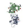
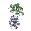
 Movie
Movie Controller


+ Open data

- Basic information
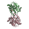
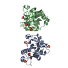
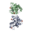
| Entry | Database: PDB / ID: 1zoo | ||||||
|---|---|---|---|---|---|---|---|
| Title | CD11A I-DOMAIN WITH BOUND MAGNESIUM ION | ||||||
 Components Components | LEUKOCYTE ADHESION GLYCOPROTEIN | ||||||
 Keywords Keywords | CELL ADHESION / INTEGRIN / GLYCOPROTEIN / TRANSMEMBRANE / EXTRACELLULAR MATRIX / CYTOSKELETON | ||||||
| Function / homology |  Function and homology information Function and homology informationmemory T cell extravasation / integrin alphaL-beta2 complex / ICAM-3 receptor activity / T cell activation via T cell receptor contact with antigen bound to MHC molecule on antigen presenting cell / RUNX3 Regulates Immune Response and Cell Migration / heterophilic cell-cell adhesion / integrin complex / leukocyte cell-cell adhesion / receptor clustering / Integrin cell surface interactions ...memory T cell extravasation / integrin alphaL-beta2 complex / ICAM-3 receptor activity / T cell activation via T cell receptor contact with antigen bound to MHC molecule on antigen presenting cell / RUNX3 Regulates Immune Response and Cell Migration / heterophilic cell-cell adhesion / integrin complex / leukocyte cell-cell adhesion / receptor clustering / Integrin cell surface interactions / specific granule membrane / phagocytosis / cell adhesion molecule binding / cell-matrix adhesion / Cell surface interactions at the vascular wall / integrin-mediated signaling pathway / cell-cell adhesion / Immunoregulatory interactions between a Lymphoid and a non-Lymphoid cell / signaling receptor activity / cell adhesion / inflammatory response / Neutrophil degranulation / protein-containing complex binding / cell surface / signal transduction / extracellular exosome / membrane / metal ion binding / plasma membrane Similarity search - Function | ||||||
| Biological species |  Homo sapiens (human) Homo sapiens (human) | ||||||
| Method |  X-RAY DIFFRACTION / Resolution: 3 Å X-RAY DIFFRACTION / Resolution: 3 Å | ||||||
 Authors Authors | Leahy, D.J. / Qu, A. | ||||||
 Citation Citation | Journal: Structure / Year: 1996 Title: The role of the divalent cation in the structure of the I domain from the CD11a/CD18 integrin. Authors: Qu, A. / Leahy, D.J. #1: Journal: Proc.Natl.Acad.Sci.USA / Year: 1995Title: Crystal Structure of the I-Domain from the Cd11A/Cd18 (Lfa-1, Alpha L Beta 2) Integrin Authors: Qu, A. / Leahy, D.J. | ||||||
| History |
|
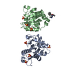
- Structure visualization
| Structure viewer | Molecule: MolmilJmol/JSmol |
|---|
- Downloads & links
-Download
| PDBx/mmCIF format | 1zoo.cif.gz | 96.3 KB | Display | PDBx/mmCIF format |
|---|---|---|---|---|
| PDB format | pdb1zoo.ent.gz | 74.3 KB | Display | PDB format |
| PDBx/mmJSON format | 1zoo.json.gz | Tree view | PDBx/mmJSON format | |
| Others |  Other downloads Other downloads |
-Validation report
| Arichive directory | https://data.pdbj.org/pub/pdb/validation_reports/zo/1zooftp://data.pdbj.org/pub/pdb/validation_reports/zo/1zoo | HTTPS FTP |
|---|
-Related structure data
| Related structure data |  1zonC  1zopSC S: Starting model for refinement C: citing same article ( |
|---|---|
| Similar structure data |


-Links
 PDBj
PDBj


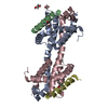
- Assembly
| Deposited unit | 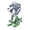
| ||||||||
|---|---|---|---|---|---|---|---|---|---|
| 1 |
| ||||||||
| Unit cell |
|
-Components
| #1: Protein | Mass: 21323.494 Da / Num. of mol.: 2 / Fragment: I-DOMAIN FRAGMENT OF LFA-1 / Mutation: W189R, MIGHT BE ISOFORM Source method: isolated from a genetically manipulated source Source: (gene. exp.) Homo sapiens (human) / Description: THE PROTEIN WAS REFOLDED FROM 7M UREA / Cell line: 293 / Gene: PCR PRODUCT / Plasmid: PET11C / Gene (production host): PCR PRODUCT / Production host:  #2: Chemical |   Mass: 24.305 Da / Num. of mol.: 2 / Source method: obtained synthetically / Formula: Mg Mass: 24.305 Da / Num. of mol.: 2 / Source method: obtained synthetically / Formula: Mg#3: Chemical |   Mass: 35.453 Da / Num. of mol.: 2 / Source method: obtained synthetically / Formula: Cl Mass: 35.453 Da / Num. of mol.: 2 / Source method: obtained synthetically / Formula: Cl#4: Water | ChemComp-HOH / |  Mass: 18.015 Da / Num. of mol.: 4 / Source method: isolated from a natural source / Formula: H2O Mass: 18.015 Da / Num. of mol.: 4 / Source method: isolated from a natural source / Formula: H2O |
|---|
-Experimental details
-Experiment
| Experiment | Method: X-RAY DIFFRACTION / Number of used crystals: 1 |
|---|
- Sample preparation
| Crystal | Density Matthews: 2.3 Å3/Da / Density % sol: 46.5 % | ||||||||||||||||||||||||||||||||||||||||||||||||
|---|---|---|---|---|---|---|---|---|---|---|---|---|---|---|---|---|---|---|---|---|---|---|---|---|---|---|---|---|---|---|---|---|---|---|---|---|---|---|---|---|---|---|---|---|---|---|---|---|---|
| Crystal grow | pH: 5.2 / Details: pH 5.2 | ||||||||||||||||||||||||||||||||||||||||||||||||
| Crystal grow | *PLUS Method: vapor diffusion, hanging drop | ||||||||||||||||||||||||||||||||||||||||||||||||
| Components of the solutions | *PLUS
|
-Data collection
| Diffraction | Mean temperature: 100 K |
|---|---|
| Diffraction source | Source: ROTATING ANODE / Wavelength: 1.5418 |
| Detector | Type: RIGAKU RAXIS IIC / Detector: IMAGE PLATE / Date: Jan 1, 1996 |
| Radiation | Monochromatic (M) / Laue (L): M / Scattering type: x-ray |
| Radiation wavelength | Wavelength: 1.5418 Å / Relative weight: 1 |
| Reflection | Resolution: 3→30 Å / Num. obs: 7748 / % possible obs: 96 % / Redundancy: 13.2 % / Rmerge(I) obs: 0.121 / Net I/σ(I): 19.2 |
- Processing
| Software |
| ||||||||||||||||||||||||||||||||||||||||||||||||||||||||||||
|---|---|---|---|---|---|---|---|---|---|---|---|---|---|---|---|---|---|---|---|---|---|---|---|---|---|---|---|---|---|---|---|---|---|---|---|---|---|---|---|---|---|---|---|---|---|---|---|---|---|---|---|---|---|---|---|---|---|---|---|---|---|
| Refinement | Starting model: 1ZOP Resolution: 3→8 Å / σ(F): 2
| ||||||||||||||||||||||||||||||||||||||||||||||||||||||||||||
| Displacement parameters | Biso mean: 11.48 Å2 | ||||||||||||||||||||||||||||||||||||||||||||||||||||||||||||
| Refinement step | Cycle: LAST / Resolution: 3→8 Å
| ||||||||||||||||||||||||||||||||||||||||||||||||||||||||||||
| Refine LS restraints |
| ||||||||||||||||||||||||||||||||||||||||||||||||||||||||||||
| Software | *PLUS Name: X-PLOR / Classification: refinement | ||||||||||||||||||||||||||||||||||||||||||||||||||||||||||||
| Refinement | *PLUS Rfactor all: 0.292 | ||||||||||||||||||||||||||||||||||||||||||||||||||||||||||||
| Solvent computation | *PLUS | ||||||||||||||||||||||||||||||||||||||||||||||||||||||||||||
| Displacement parameters | *PLUS |
