 Movie
Movie Controller

[English] 日本語
 Yorodumi
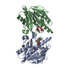
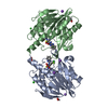
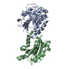
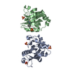
- PDB-1gka: The molecular basis of the coloration mechanism in lobster shell.... -
+ Open data

- Basic information
| Entry | Database: PDB / ID: 1gka | ||||||
|---|---|---|---|---|---|---|---|
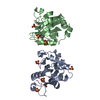
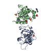
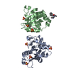
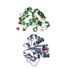
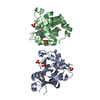
| Title | The molecular basis of the coloration mechanism in lobster shell. beta-crustacyanin at 3.2 A resolution | ||||||
 Components Components | (CRUSTACYANIN ...) x 2 | ||||||
 Keywords Keywords | LIPOCALIN / CRUSTACYANIN / LOBSTER / ASTAXANTHIN / BATHOCHROMIC / COLORATION | ||||||
| Function / homology |  Function and homology information Function and homology informationpigment binding / response to reactive oxygen species / lipid metabolic process / extracellular region / cytoplasm Similarity search - Function | ||||||
| Biological species |  HOMARUS GAMMARUS (European lobster) HOMARUS GAMMARUS (European lobster) | ||||||
| Method |  X-RAY DIFFRACTION / SYNCHROTRON / MOLECULAR REPLACEMENT / Resolution: 3.23 Å X-RAY DIFFRACTION / SYNCHROTRON / MOLECULAR REPLACEMENT / Resolution: 3.23 Å | ||||||
 Authors Authors | Cianci, M. / Rizkallah, P.J. / Olczak, A. / Raftery, J. / Chayen, N.E. / Zagalsky, P.F. / Helliwell, J.R. | ||||||
 Citation Citation | Journal: Proc.Natl.Acad.Sci.USA / Year: 2002 Title: The Molecular Basis of the Coloration Mechanism in Lobster Shell: Beta -Crustacyanin at 3.2-A Resolution Authors: Cianci, M. / Rizkallah, P. / Olczak, A. / Raftery, J. / Chayen, N. / Zagalsky, P. / Helliwell, J. | ||||||
| History |
| ||||||
| Remark 700 | SHEET DETERMINATION METHOD: DSSP THE SHEETS PRESENTED AS "AA" IN EACH CHAIN ON SHEET RECORDS BELOW ... SHEET DETERMINATION METHOD: DSSP THE SHEETS PRESENTED AS "AA" IN EACH CHAIN ON SHEET RECORDS BELOW IS ACTUALLY AN 9-STRANDED BARREL THIS IS REPRESENTED BY A 10-STRANDED SHEET IN WHICH THE FIRST AND LAST STRANDS ARE IDENTICAL. REMARK 900 |
- Structure visualization
| Structure viewer | Molecule: MolmilJmol/JSmol |
|---|
- Downloads & links
-Download
| PDBx/mmCIF format | 1gka.cif.gz | 89.7 KB | Display | PDBx/mmCIF format |
|---|---|---|---|---|
| PDB format | pdb1gka.ent.gz | 67.9 KB | Display | PDB format |
| PDBx/mmJSON format | 1gka.json.gz | Tree view | PDBx/mmJSON format | |
| Others |  Other downloads Other downloads |
-Validation report
| Arichive directory | https://data.pdbj.org/pub/pdb/validation_reports/gk/1gkaftp://data.pdbj.org/pub/pdb/validation_reports/gk/1gka | HTTPS FTP |
|---|
-Related structure data
| Related structure data |  1h91S S: Starting model for refinement |
|---|---|
| Similar structure data |

-Links
 PDBj
PDBj

- Assembly
| Deposited unit | 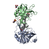
| ||||||||
|---|---|---|---|---|---|---|---|---|---|
| 1 |
| ||||||||
| Unit cell |
|
-Components
-CRUSTACYANIN ... , 2 types, 2 molecules AB
| #1: Protein | Mass: 20558.029 Da / Num. of mol.: 1 / Source method: isolated from a natural source / Source: (natural) HOMARUS GAMMARUS (European lobster) / Tissue: CARAPACE / References: UniProt: P58989*PLUS |
|---|---|
| #2: Protein | Mass: 19688.822 Da / Num. of mol.: 1 / Source method: isolated from a natural source / Source: (natural) HOMARUS GAMMARUS (European lobster) / Tissue: CARAPACE / References: UniProt: P80007 |
-Non-polymers , 5 types, 35 molecules 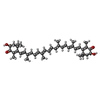


| #3: Chemical |  Mass: 596.838 Da / Num. of mol.: 2 / Source method: obtained synthetically / Formula: C40H52O4 Mass: 596.838 Da / Num. of mol.: 2 / Source method: obtained synthetically / Formula: C40H52O4#4: Chemical | ChemComp-D12 / |  Mass: 170.335 Da / Num. of mol.: 1 / Source method: obtained synthetically / Formula: C12H26 Mass: 170.335 Da / Num. of mol.: 1 / Source method: obtained synthetically / Formula: C12H26#5: Chemical | ChemComp-TRS / |  Mass: 122.143 Da / Num. of mol.: 1 / Source method: obtained synthetically / Formula: C4H12NO3 / Comment: pH buffer*YM Mass: 122.143 Da / Num. of mol.: 1 / Source method: obtained synthetically / Formula: C4H12NO3 / Comment: pH buffer*YM#6: Chemical | ChemComp-EPE / |  Mass: 238.305 Da / Num. of mol.: 1 / Source method: obtained synthetically / Formula: C8H18N2O4S / Comment: pH buffer*YM Mass: 238.305 Da / Num. of mol.: 1 / Source method: obtained synthetically / Formula: C8H18N2O4S / Comment: pH buffer*YM#7: Water | ChemComp-HOH / | Mass: 18.015 Da / Num. of mol.: 30 / Source method: isolated from a natural source / Formula: H2O |
|---|
-Details
| Compound details | THIS BETA CRUSTACYANIN IS RESPONSIBLE FOR BINDING THE CAROTENOID ASTAXANTHIN THAT PROVIDES THE ...THIS BETA CRUSTACYAN |
|---|---|
| Has protein modification | Y |
-Experimental details
-Experiment
| Experiment | Method: X-RAY DIFFRACTION / Number of used crystals: 1 |
|---|
- Sample preparation
| Crystal | Density Matthews: 7.3 Å3/Da / Density % sol: 78.89 % | ||||||||||||||||||||||||||||||
|---|---|---|---|---|---|---|---|---|---|---|---|---|---|---|---|---|---|---|---|---|---|---|---|---|---|---|---|---|---|---|---|
| Crystal grow | pH: 7.7 / Details: pH 7.70 | ||||||||||||||||||||||||||||||
| Crystal grow | *PLUS Temperature: 4 ℃ / pH: 8 / Method: vapor diffusion | ||||||||||||||||||||||||||||||
| Components of the solutions | *PLUS
|
-Data collection
| Diffraction | Mean temperature: 100 K |
|---|---|
| Diffraction source | Source: SYNCHROTRON / Site: SRS  / Beamline: PX14.1 / Wavelength: 1.488 / Beamline: PX14.1 / Wavelength: 1.488 |
| Detector | Date: Nov 15, 2000 |
| Radiation | Protocol: SINGLE WAVELENGTH / Monochromatic (M) / Laue (L): M / Scattering type: x-ray |
| Radiation wavelength | Wavelength: 1.488 Å / Relative weight: 1 |
| Reflection | Resolution: 3→100 Å / Num. obs: 24504 / % possible obs: 99.7 % / Observed criterion σ(I): 0 / Redundancy: 7 % / Rmerge(I) obs: 0.124 / Net I/σ(I): 15.5 |
| Reflection shell | Resolution: 3.23→3.38 Å / Rmerge(I) obs: 0.35 / Mean I/σ(I) obs: 5.5 / % possible all: 100 |
| Reflection | *PLUS Highest resolution: 3 Å / Lowest resolution: 100 Å / Num. measured all: 171943 |
| Reflection shell | *PLUS % possible obs: 100 % / Rmerge(I) obs: 0.35 |
- Processing
| Software |
| ||||||||||||||||||||||||||||||||||||||||||||||||||||||||||||
|---|---|---|---|---|---|---|---|---|---|---|---|---|---|---|---|---|---|---|---|---|---|---|---|---|---|---|---|---|---|---|---|---|---|---|---|---|---|---|---|---|---|---|---|---|---|---|---|---|---|---|---|---|---|---|---|---|---|---|---|---|---|
| Refinement | Method to determine structure: MOLECULAR REPLACEMENT Starting model: 1H91 Resolution: 3.23→100 Å / σ(F): 0
| ||||||||||||||||||||||||||||||||||||||||||||||||||||||||||||
| Refine analyze |
| ||||||||||||||||||||||||||||||||||||||||||||||||||||||||||||
| Refinement step | Cycle: LAST / Resolution: 3.23→100 Å
| ||||||||||||||||||||||||||||||||||||||||||||||||||||||||||||
| Refine LS restraints |
| ||||||||||||||||||||||||||||||||||||||||||||||||||||||||||||
| LS refinement shell | Resolution: 3.23→3.35 Å / Total num. of bins used: 10
| ||||||||||||||||||||||||||||||||||||||||||||||||||||||||||||
| Refinement | *PLUS Lowest resolution: 100 Å / Rfactor Rfree: 0.251 / Rfactor Rwork: 0.213 | ||||||||||||||||||||||||||||||||||||||||||||||||||||||||||||
| Solvent computation | *PLUS | ||||||||||||||||||||||||||||||||||||||||||||||||||||||||||||
| Displacement parameters | *PLUS | ||||||||||||||||||||||||||||||||||||||||||||||||||||||||||||
| Refine LS restraints | *PLUS
|
