 Movie
Movie Controller

[English] 日本語
 Yorodumi
- PDB-7cz0: Crystal structure of a thermostable green fluorescent protein (TG... -
+ Open data

- Basic information
| Entry | Database: PDB / ID: 7cz0 | ||||||||||||
|---|---|---|---|---|---|---|---|---|---|---|---|---|---|
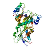
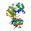
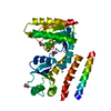
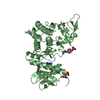
| Title | Crystal structure of a thermostable green fluorescent protein (TGP) with a synthetic nanobody (Sb92) | ||||||||||||
 Components Components |
| ||||||||||||
 Keywords Keywords | FLUORESCENT PROTEIN / complex / GFP / nanobody / single-chain antibody / sybody / synthetic antibody / TGP / thermostable green fluorescent protein | ||||||||||||
| Function / homology | Immunoglobulins / Immunoglobulin-like / Sandwich / Mainly Beta / ACETATE ION / CACODYLATE ION / CACODYLIC ACID Function and homology information Function and homology information | ||||||||||||
| Biological species |  Galaxea fascicularis (invertebrata) Galaxea fascicularis (invertebrata)synthetic construct (others) | ||||||||||||
| Method |  X-RAY DIFFRACTION / SYNCHROTRON / MOLECULAR REPLACEMENT / Resolution: 2.77 Å X-RAY DIFFRACTION / SYNCHROTRON / MOLECULAR REPLACEMENT / Resolution: 2.77 Å | ||||||||||||
 Authors Authors | Cai, H. / Yao, H. / Li, T. / Hutter, C. / Tang, Y. / Li, Y. / Seeger, M. / Li, D. | ||||||||||||
| Funding support |  China, 1items China, 1items
| ||||||||||||
 Citation Citation | Journal: To Be Published Title: An improved fluorescent protein tag and its nanobodies for membrane protein expression, stability assay, and purification Authors: Cai, H. / Yao, H. / Li, T. / Hutter, C. / Tang, Y. / Li, Y. / Seeger, M. / Li, D. | ||||||||||||
| History |
|
- Structure visualization
| Structure viewer | Molecule: MolmilJmol/JSmol |
|---|
- Downloads & links
-Download
| PDBx/mmCIF format | 7cz0.cif.gz | 334.3 KB | Display | PDBx/mmCIF format |
|---|---|---|---|---|
| PDB format | pdb7cz0.ent.gz | 219.1 KB | Display | PDB format |
| PDBx/mmJSON format | 7cz0.json.gz | Tree view | PDBx/mmJSON format | |
| Others |  Other downloads Other downloads |
-Validation report
| Arichive directory | https://data.pdbj.org/pub/pdb/validation_reports/cz/7cz0ftp://data.pdbj.org/pub/pdb/validation_reports/cz/7cz0 | HTTPS FTP |
|---|
-Related structure data
| Related structure data |  6lz2S S: Starting model for refinement |
|---|---|
| Similar structure data |


-Links
 PDBj
PDBj


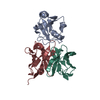
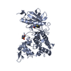
- Assembly
| Deposited unit | 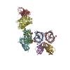
| ||||||||||||
|---|---|---|---|---|---|---|---|---|---|---|---|---|---|
| 1 | 
| ||||||||||||
| 2 | 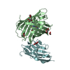
| ||||||||||||
| 3 | 
| ||||||||||||
| 4 | 
| ||||||||||||
| Unit cell |
|
-Components
-Protein / Antibody , 2 types, 8 molecules ABCDEFGH
| #1: Protein | Mass: 26699.957 Da / Num. of mol.: 4 Source method: isolated from a genetically manipulated source Details: Uniprot A8CLT2 / Source: (gene. exp.) Galaxea fascicularis (invertebrata) / Plasmid: pEC / Details (production host): pET-based vector / Production host:  #2: Antibody | Mass: 15811.404 Da / Num. of mol.: 4 Source method: isolated from a genetically manipulated source Source: (gene. exp.) synthetic construct (others) / Plasmid: pEC / Details (production host): pET-based vector / Production host: |
|---|
-Non-polymers , 4 types, 15 molecules 
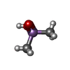


| #3: Chemical | ChemComp-GOL /  Mass: 92.094 Da / Num. of mol.: 11 / Source method: obtained synthetically / Formula: C3H8O3 Mass: 92.094 Da / Num. of mol.: 11 / Source method: obtained synthetically / Formula: C3H8O3#4: Chemical | ChemComp-CAD / |  Mass: 137.997 Da / Num. of mol.: 1 / Source method: obtained synthetically / Formula: C2H7AsO2 Mass: 137.997 Da / Num. of mol.: 1 / Source method: obtained synthetically / Formula: C2H7AsO2#5: Chemical |  Mass: 59.044 Da / Num. of mol.: 2 / Source method: obtained synthetically / Formula: C2H3O2 Mass: 59.044 Da / Num. of mol.: 2 / Source method: obtained synthetically / Formula: C2H3O2#6: Chemical | ChemComp-CAC / |  Mass: 136.989 Da / Num. of mol.: 1 / Source method: obtained synthetically / Formula: C2H6AsO2 Mass: 136.989 Da / Num. of mol.: 1 / Source method: obtained synthetically / Formula: C2H6AsO2 |
|---|
-Details
| Has ligand of interest | Y |
|---|---|
| Has protein modification | Y |
-Experimental details
-Experiment
| Experiment | Method: X-RAY DIFFRACTION / Number of used crystals: 1 |
|---|
- Sample preparation
| Crystal | Density Matthews: 3.12 Å3/Da / Density % sol: 64.93 % |
|---|---|
| Crystal grow | Temperature: 293 K / Method: vapor diffusion, sitting drop / pH: 6.5 Details: 14.4 %(w/v) PEG 8000, 160mM calcium acetate, 20% (v/v) glycerol, 80mM sodium cacodylate / HCl pH 6.5 |
-Data collection
| Diffraction | Mean temperature: 100 K / Serial crystal experiment: N |
|---|---|
| Diffraction source | Source: SYNCHROTRON / Site: SSRF / Beamline: BL18U1 / Wavelength: 0.9793 Å |
| Detector | Type: PSI PILATUS 6M / Detector: PIXEL / Date: Mar 12, 2020 |
| Radiation | Protocol: SINGLE WAVELENGTH / Monochromatic (M) / Laue (L): M / Scattering type: x-ray |
| Radiation wavelength | Wavelength: 0.9793 Å / Relative weight: 1 |
| Reflection | Resolution: 2.77→47.62 Å / Num. obs: 54942 / % possible obs: 99.9 % / Redundancy: 6.7 % / Biso Wilson estimate: 56.03 Å2 / CC1/2: 0.997 / Rmerge(I) obs: 0.148 / Rpim(I) all: 0.062 / Net I/σ(I): 9.2 |
| Reflection shell | Resolution: 2.77→2.85 Å / Redundancy: 7 % / Rmerge(I) obs: 1.147 / Mean I/σ(I) obs: 1.7 / Num. unique obs: 4418 / CC1/2: 0.78 / Rpim(I) all: 0.651 / % possible all: 99.9 |
- Processing
| Software |
| ||||||||||||||||||||||||||||||||||||||||||||||||||||||||||||||||||||||||||||||||||||||||||||||||||||||||||||||||||||||||||||||||||||||||||||
|---|---|---|---|---|---|---|---|---|---|---|---|---|---|---|---|---|---|---|---|---|---|---|---|---|---|---|---|---|---|---|---|---|---|---|---|---|---|---|---|---|---|---|---|---|---|---|---|---|---|---|---|---|---|---|---|---|---|---|---|---|---|---|---|---|---|---|---|---|---|---|---|---|---|---|---|---|---|---|---|---|---|---|---|---|---|---|---|---|---|---|---|---|---|---|---|---|---|---|---|---|---|---|---|---|---|---|---|---|---|---|---|---|---|---|---|---|---|---|---|---|---|---|---|---|---|---|---|---|---|---|---|---|---|---|---|---|---|---|---|---|---|
| Refinement | Method to determine structure: MOLECULAR REPLACEMENT Starting model: 6LZ2 Resolution: 2.77→47.62 Å / SU ML: 0.3827 / Cross valid method: FREE R-VALUE / σ(F): 1.34 / Phase error: 29.3841 Stereochemistry target values: GeoStd + Monomer Library + CDL v1.2
| ||||||||||||||||||||||||||||||||||||||||||||||||||||||||||||||||||||||||||||||||||||||||||||||||||||||||||||||||||||||||||||||||||||||||||||
| Solvent computation | Shrinkage radii: 0.9 Å / VDW probe radii: 1.11 Å / Solvent model: FLAT BULK SOLVENT MODEL | ||||||||||||||||||||||||||||||||||||||||||||||||||||||||||||||||||||||||||||||||||||||||||||||||||||||||||||||||||||||||||||||||||||||||||||
| Displacement parameters | Biso mean: 54.23 Å2 | ||||||||||||||||||||||||||||||||||||||||||||||||||||||||||||||||||||||||||||||||||||||||||||||||||||||||||||||||||||||||||||||||||||||||||||
| Refinement step | Cycle: LAST / Resolution: 2.77→47.62 Å
| ||||||||||||||||||||||||||||||||||||||||||||||||||||||||||||||||||||||||||||||||||||||||||||||||||||||||||||||||||||||||||||||||||||||||||||
| Refine LS restraints |
| ||||||||||||||||||||||||||||||||||||||||||||||||||||||||||||||||||||||||||||||||||||||||||||||||||||||||||||||||||||||||||||||||||||||||||||
| LS refinement shell |
|
