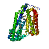
 Movie
Movie Controller


+ Open data

- Basic information
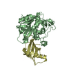
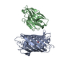
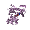
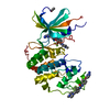
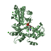
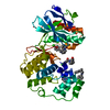
| Entry | Database: PDB / ID: 3kse | ||||||
|---|---|---|---|---|---|---|---|
| Title | Unreduced cathepsin L in complex with stefin A | ||||||
 Components Components |
| ||||||
 Keywords Keywords | Hydrolase/Hydrolase inhibitor / cathepsin / protease-inhibitor complex / stefin / cystatin / papain-like / cysteine protease / Hydrolase / Lysosome / Protease / Thiol protease / Zymogen / Thiol protease inhibitor / Hydrolase-Hydrolase inhibitor complex | ||||||
| Function / homology |  Function and homology information Function and homology informationenkephalin processing / cathepsin L / CD4-positive, alpha-beta T cell lineage commitment / peptidase inhibitor complex / macrophage apoptotic process / peptide cross-linking / chromaffin granule / antigen processing and presentation of peptide antigen / elastin catabolic process / Formation of the cornified envelope ...enkephalin processing / cathepsin L / CD4-positive, alpha-beta T cell lineage commitment / peptidase inhibitor complex / macrophage apoptotic process / peptide cross-linking / chromaffin granule / antigen processing and presentation of peptide antigen / elastin catabolic process / Formation of the cornified envelope / HS-GAG degradation / RUNX1 regulates transcription of genes involved in differentiation of keratinocytes / cornified envelope / endolysosome lumen / cellular response to thyroid hormone stimulus / Trafficking and processing of endosomal TLR / proteoglycan binding / Assembly of collagen fibrils and other multimeric structures / zymogen activation / antigen processing and presentation / Collagen degradation / protein autoprocessing / collagen catabolic process / fibronectin binding / serpin family protein binding / cysteine-type endopeptidase inhibitor activity / keratinocyte differentiation / Degradation of the extracellular matrix / collagen binding / receptor-mediated endocytosis of virus by host cell / negative regulation of proteolysis / Attachment and Entry / multivesicular body / endocytic vesicle lumen / cysteine-type peptidase activity / MHC class II antigen presentation / lysosomal lumen / : / Endosomal/Vacuolar pathway / cell-cell adhesion / Degradation of CDH1 / antigen processing and presentation of exogenous peptide antigen via MHC class II / extracellular matrix / protease binding / histone binding / adaptive immune response / Attachment and Entry / lysosome / apical plasma membrane / fusion of virus membrane with host plasma membrane / cysteine-type endopeptidase activity / fusion of virus membrane with host endosome membrane / symbiont entry into host cell / Golgi apparatus / proteolysis / : / extracellular exosome / extracellular region / nucleoplasm / nucleus / plasma membrane / cytoplasm / cytosol Similarity search - Function | ||||||
| Biological species |  Homo sapiens (human) Homo sapiens (human) | ||||||
| Method |  X-RAY DIFFRACTION / SYNCHROTRON / MOLECULAR REPLACEMENT / Resolution: 1.71 Å X-RAY DIFFRACTION / SYNCHROTRON / MOLECULAR REPLACEMENT / Resolution: 1.71 Å | ||||||
 Authors Authors | Renko, M. / Turk, D. | ||||||
 Citation Citation | Journal: To be Published Title: Unreduced cathepsin L in complex with stefin A Authors: Renko, M. / Turk, D. | ||||||
| History |
|
- Structure visualization
| Structure viewer | Molecule: MolmilJmol/JSmol |
|---|
- Downloads & links
-Download
| PDBx/mmCIF format | 3kse.cif.gz | 220.8 KB | Display | PDBx/mmCIF format |
|---|---|---|---|---|
| PDB format | pdb3kse.ent.gz | 174.7 KB | Display | PDB format |
| PDBx/mmJSON format | 3kse.json.gz | Tree view | PDBx/mmJSON format | |
| Others |  Other downloads Other downloads |
-Validation report
| Arichive directory | https://data.pdbj.org/pub/pdb/validation_reports/ks/3kseftp://data.pdbj.org/pub/pdb/validation_reports/ks/3kse | HTTPS FTP |
|---|
-Related structure data
| Related structure data | 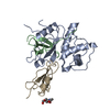 1icfS  3kfqS  3a9n S: Starting model for refinement |
|---|---|
| Similar structure data |


-Links
 PDBj
PDBj


- Assembly
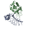
| Deposited unit | 
| ||||||||
|---|---|---|---|---|---|---|---|---|---|
| 1 | 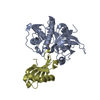
| ||||||||
| 2 | 
| ||||||||
| 3 | 
| ||||||||
| Unit cell |
|
-Components
| #1: Protein | Mass: 24208.754 Da / Num. of mol.: 3 / Mutation: T110A,N179D Source method: isolated from a genetically manipulated source Source: (gene. exp.) Homo sapiens (human) / Plasmid: pPIC9 / Production host:  Pichia Pastoris (fungus) / Strain (production host): GS115 / References: UniProt: P07711, cathepsin L Pichia Pastoris (fungus) / Strain (production host): GS115 / References: UniProt: P07711, cathepsin L#2: Protein | Mass: 11020.464 Da / Num. of mol.: 3 Source method: isolated from a genetically manipulated source Source: (gene. exp.) Homo sapiens (human) / Plasmid: pET3a / Production host:  #3: Water | ChemComp-HOH / |  Mass: 18.015 Da / Num. of mol.: 1143 / Source method: isolated from a natural source / Formula: H2O Mass: 18.015 Da / Num. of mol.: 1143 / Source method: isolated from a natural source / Formula: H2OSequence details | CATHEPSIN L WAS BLOCKED WITH MMTS (METHYL METHANETHIOSULFONATE), LEAVING -S-CH3 ATOMS ON ACTIVE ...CATHEPSIN L WAS BLOCKED WITH MMTS (METHYL METHANETHI | |
|---|
-Experimental details
-Experiment
| Experiment | Method: X-RAY DIFFRACTION / Number of used crystals: 1 |
|---|
- Sample preparation
| Crystal | Density Matthews: 1.99 Å3/Da / Density % sol: 38.31 % |
|---|---|
| Crystal grow | Temperature: 293 K / Method: vapor diffusion, sitting drop / pH: 7 Details: 0.1M Tris-HCl, 16% PEG3000, pH 7.0, VAPOR DIFFUSION, SITTING DROP, temperature 293K |
-Data collection
| Diffraction | Mean temperature: 100 K |
|---|---|
| Diffraction source | Source: SYNCHROTRON / Site: ELETTRA  / Beamline: 5.2R / Wavelength: 1 Å / Beamline: 5.2R / Wavelength: 1 Å |
| Detector | Type: MAR CCD 165 mm / Detector: CCD / Date: Aug 20, 2008 / Details: Collimating and focusing, Pt-coated mirrors |
| Radiation | Monochromator: Double Crystal Si111 or White Beam / Protocol: SINGLE WAVELENGTH / Monochromatic (M) / Laue (L): M / Scattering type: x-ray |
| Radiation wavelength | Wavelength: 1 Å / Relative weight: 1 |
| Reflection | Resolution: 1.69→50 Å / Num. all: 90358 / Num. obs: 86021 / % possible obs: 95.2 % / Observed criterion σ(F): 1 / Observed criterion σ(I): 1 / Redundancy: 2 % / Rmerge(I) obs: 0.04 / Net I/σ(I): 13.3 |
| Reflection shell | Resolution: 1.69→1.72 Å / Redundancy: 1.3 % / Rmerge(I) obs: 0.273 / Num. unique all: 2830 / % possible all: 62.7 |
- Processing
| Software |
| |||||||||||||||||||||||||
|---|---|---|---|---|---|---|---|---|---|---|---|---|---|---|---|---|---|---|---|---|---|---|---|---|---|---|
| Refinement | Method to determine structure: MOLECULAR REPLACEMENT Starting model: 3KFQ, 1ICF Resolution: 1.71→15.33 Å / Cross valid method: THROUGHOUT / σ(F): 0 / Stereochemistry target values: MAXIMUM LIKELIHOOD Details: HYDROGENS HAVE BEEN ADDED IN THE RIDING POSITIONS U VALUES: REFINED INDIVIDUALLY
| |||||||||||||||||||||||||
| Displacement parameters | Biso mean: 18.15 Å2
| |||||||||||||||||||||||||
| Refinement step | Cycle: LAST / Resolution: 1.71→15.33 Å
| |||||||||||||||||||||||||
| Refine LS restraints |
| |||||||||||||||||||||||||
| LS refinement shell | Resolution: 1.706→1.75 Å
|
