 Movie
Movie Controller

[English] 日本語
 Yorodumi
- PDB-7cxd: Xray structure of rat Galectin-3 CRD in complex with TD-139 belon... -
+ Open data

- Basic information
| Entry | Database: PDB / ID: 7cxd | ||||||
|---|---|---|---|---|---|---|---|
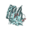
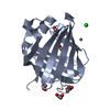
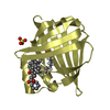
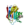
| Title | Xray structure of rat Galectin-3 CRD in complex with TD-139 belonging to P121 space group | ||||||
 Components Components | Galectin-3 | ||||||
 Keywords Keywords | SUGAR BINDING PROTEIN / Beta-galactose binding protein / CARBOHYDRATE / TD-139 | ||||||
| Function / homology |  Function and homology information Function and homology informationnegative regulation of cell proliferation in bone marrow / advanced glycation end-product receptor activity / positive regulation of serotonin secretion / response to quercetin / negative regulation of immunological synapse formation / cornified envelope / negative regulation of T cell activation via T cell receptor contact with antigen bound to MHC molecule on antigen presenting cell / disaccharide binding / regulation of T cell apoptotic process / mononuclear cell migration ...negative regulation of cell proliferation in bone marrow / advanced glycation end-product receptor activity / positive regulation of serotonin secretion / response to quercetin / negative regulation of immunological synapse formation / cornified envelope / negative regulation of T cell activation via T cell receptor contact with antigen bound to MHC molecule on antigen presenting cell / disaccharide binding / regulation of T cell apoptotic process / mononuclear cell migration / negative regulation of endocytosis / positive regulation of mononuclear cell migration / IgE binding / eosinophil chemotaxis / monosaccharide binding / Fc-gamma receptor I complex binding / regulation of extrinsic apoptotic signaling pathway via death domain receptors / Neutrophil degranulation / positive chemotaxis / regulation of T cell proliferation / chemoattractant activity / positive regulation of calcium ion import / macrophage chemotaxis / monocyte chemotaxis / immunological synapse / negative regulation of T cell receptor signaling pathway / glial cell projection / neutrophil chemotaxis / extracellular matrix organization / epithelial cell differentiation / laminin binding / RNA splicing / negative regulation of extrinsic apoptotic signaling pathway / skeletal system development / positive regulation of protein localization to plasma membrane / spliceosomal complex / molecular condensate scaffold activity / positive regulation of protein-containing complex assembly / positive regulation of angiogenesis / mRNA processing / carbohydrate binding / extracellular matrix / protein phosphatase binding / mitochondrial inner membrane / external side of plasma membrane / innate immune response / positive regulation of cell population proliferation / negative regulation of apoptotic process / cell surface / : / nucleus / plasma membrane / cytoplasm Similarity search - Function | ||||||
| Biological species |  | ||||||
| Method |  X-RAY DIFFRACTION / SYNCHROTRON / MOLECULAR REPLACEMENT / Resolution: 1.71 Å X-RAY DIFFRACTION / SYNCHROTRON / MOLECULAR REPLACEMENT / Resolution: 1.71 Å | ||||||
 Authors Authors | Kumar, A. | ||||||
 Citation Citation | Journal: Glycobiology / Year: 2021 Title: Molecular mechanism of interspecies differences in the binding affinity of TD139 to Galectin-3. Authors: Kumar, A. / Paul, M. / Panda, M. / Jayaram, S. / Kalidindi, N. / Sale, H. / Vetrichelvan, M. / Gupta, A. / Mathur, A. / Beno, B. / Regueiro-Ren, A. / Cheng, D. / Ramarao, M. / Ghosh, K. | ||||||
| History |
|
- Structure visualization
| Structure viewer | Molecule: MolmilJmol/JSmol |
|---|
- Downloads & links
-Download
| PDBx/mmCIF format | 7cxd.cif.gz | 135.2 KB | Display | PDBx/mmCIF format |
|---|---|---|---|---|
| PDB format | pdb7cxd.ent.gz | 104.5 KB | Display | PDB format |
| PDBx/mmJSON format | 7cxd.json.gz | Tree view | PDBx/mmJSON format | |
| Others |  Other downloads Other downloads |
-Validation report
| Arichive directory | https://data.pdbj.org/pub/pdb/validation_reports/cx/7cxdftp://data.pdbj.org/pub/pdb/validation_reports/cx/7cxd | HTTPS FTP |
|---|
-Related structure data
| Related structure data |  7cxaSC 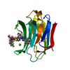 7cxbC 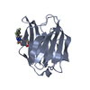 7cxcC S: Starting model for refinement C: citing same article ( |
|---|---|
| Similar structure data |


-Links
 PDBj
PDBj


- Assembly
| Deposited unit | 
| ||||||||
|---|---|---|---|---|---|---|---|---|---|
| 1 | 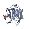
| ||||||||
| 2 | 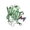
| ||||||||
| 3 | 
| ||||||||
| 4 | 
| ||||||||
| Unit cell |
|
-Components
| #1: Protein | Mass: 16400.873 Da / Num. of mol.: 4 / Fragment: Carbohydrare Recognition Domain Source method: isolated from a genetically manipulated source Source: (gene. exp.)  #2: Chemical | ChemComp-BR /   Mass: 79.904 Da / Num. of mol.: 5 / Source method: obtained synthetically / Formula: Br Mass: 79.904 Da / Num. of mol.: 5 / Source method: obtained synthetically / Formula: Br#3: Chemical | ChemComp-TD2 /   Mass: 648.635 Da / Num. of mol.: 4 / Source method: obtained synthetically / Formula: C28H30F2N6O8S / Feature type: SUBJECT OF INVESTIGATION Mass: 648.635 Da / Num. of mol.: 4 / Source method: obtained synthetically / Formula: C28H30F2N6O8S / Feature type: SUBJECT OF INVESTIGATION#4: Water | ChemComp-HOH / |  Mass: 18.015 Da / Num. of mol.: 266 / Source method: isolated from a natural source / Formula: H2O Mass: 18.015 Da / Num. of mol.: 266 / Source method: isolated from a natural source / Formula: H2OHas ligand of interest | Y | |
|---|
-Experimental details
-Experiment
| Experiment | Method: X-RAY DIFFRACTION / Number of used crystals: 1 |
|---|
- Sample preparation
| Crystal | Density Matthews: 1.95 Å3/Da / Density % sol: 36.85 % |
|---|---|
| Crystal grow | Temperature: 293.15 K / Method: vapor diffusion, sitting drop Details: 24-35% PEG 4000/6000, 0.1M Tris (pH 7.5 to pH 8.5), 0.1M MgCl2, 0.4M NaSCN |
-Data collection
| Diffraction | Mean temperature: 100 K / Serial crystal experiment: N | ||||||||||||||||||||||||||||||
|---|---|---|---|---|---|---|---|---|---|---|---|---|---|---|---|---|---|---|---|---|---|---|---|---|---|---|---|---|---|---|---|
| Diffraction source | Source: SYNCHROTRON / Site: APS  / Beamline: 21-ID-D / Wavelength: 1.078 Å / Beamline: 21-ID-D / Wavelength: 1.078 Å | ||||||||||||||||||||||||||||||
| Detector | Type: DECTRIS EIGER2 X 9M / Detector: PIXEL / Date: Feb 21, 2017 | ||||||||||||||||||||||||||||||
| Radiation | Protocol: SINGLE WAVELENGTH / Monochromatic (M) / Laue (L): M / Scattering type: x-ray | ||||||||||||||||||||||||||||||
| Radiation wavelength | Wavelength: 1.078 Å / Relative weight: 1 | ||||||||||||||||||||||||||||||
| Reflection | Resolution: 1.71→61.5 Å / Num. obs: 48235 / % possible obs: 87.9 % / Redundancy: 3.6 % / Biso Wilson estimate: 27.23 Å2 / CC1/2: 0.995 / Rmerge(I) obs: 0.074 / Rpim(I) all: 0.045 / Rrim(I) all: 0.087 / Net I/σ(I): 9.3 / Num. measured all: 172697 | ||||||||||||||||||||||||||||||
| Reflection shell | Diffraction-ID: 1
|
- Processing
| Software |
| ||||||||||||||||||||||||||||||||||||||||||||||||||||||||||||||||||||||||||||||||||||||||||||||||||||||||||||
|---|---|---|---|---|---|---|---|---|---|---|---|---|---|---|---|---|---|---|---|---|---|---|---|---|---|---|---|---|---|---|---|---|---|---|---|---|---|---|---|---|---|---|---|---|---|---|---|---|---|---|---|---|---|---|---|---|---|---|---|---|---|---|---|---|---|---|---|---|---|---|---|---|---|---|---|---|---|---|---|---|---|---|---|---|---|---|---|---|---|---|---|---|---|---|---|---|---|---|---|---|---|---|---|---|---|---|---|---|---|
| Refinement | Method to determine structure: MOLECULAR REPLACEMENT Starting model: 7CXA Resolution: 1.71→41.17 Å / Cor.coef. Fo:Fc: 0.928 / Cor.coef. Fo:Fc free: 0.907 / SU R Cruickshank DPI: 0.164 / Cross valid method: THROUGHOUT / σ(F): 0 / SU R Blow DPI: 0.163 / SU Rfree Blow DPI: 0.138 / SU Rfree Cruickshank DPI: 0.139
| ||||||||||||||||||||||||||||||||||||||||||||||||||||||||||||||||||||||||||||||||||||||||||||||||||||||||||||
| Displacement parameters | Biso max: 97.06 Å2 / Biso mean: 31.73 Å2 / Biso min: 12.49 Å2
| ||||||||||||||||||||||||||||||||||||||||||||||||||||||||||||||||||||||||||||||||||||||||||||||||||||||||||||
| Refine analyze | Luzzati coordinate error obs: 0.29 Å | ||||||||||||||||||||||||||||||||||||||||||||||||||||||||||||||||||||||||||||||||||||||||||||||||||||||||||||
| Refinement step | Cycle: final / Resolution: 1.71→41.17 Å
| ||||||||||||||||||||||||||||||||||||||||||||||||||||||||||||||||||||||||||||||||||||||||||||||||||||||||||||
| Refine LS restraints |
| ||||||||||||||||||||||||||||||||||||||||||||||||||||||||||||||||||||||||||||||||||||||||||||||||||||||||||||
| LS refinement shell | Resolution: 1.71→1.72 Å / Rfactor Rfree error: 0 / Total num. of bins used: 50
|
