 Movie
Movie Controller

[English] 日本語
 Yorodumi
- PDB-7cv1: Structure of human tRNAHis guanylyltransferase (Thg1) in the pres... -
+ Open data

- Basic information
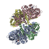
| Entry | Database: PDB / ID: 7cv1 | |||||||||
|---|---|---|---|---|---|---|---|---|---|---|
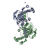
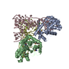
| Title | Structure of human tRNAHis guanylyltransferase (Thg1) in the presence of human mitochondrial tRNAHis | |||||||||
 Components Components | Probable tRNA(His) guanylyltransferase | |||||||||
 Keywords Keywords | TRANSFERASE / tRNA modification | |||||||||
| Function / homology |  Function and homology information Function and homology informationstress-induced mitochondrial fusion / tRNAHis guanylyltransferase / tRNA guanylyltransferase activity / transferase complex / tRNA modification in the nucleus and cytosol / tRNA modification / mitochondrial fusion / tRNA processing / nucleotidyltransferase activity / guanyl-nucleotide exchange factor activity ...stress-induced mitochondrial fusion / tRNAHis guanylyltransferase / tRNA guanylyltransferase activity / transferase complex / tRNA modification in the nucleus and cytosol / tRNA modification / mitochondrial fusion / tRNA processing / nucleotidyltransferase activity / guanyl-nucleotide exchange factor activity / response to oxidative stress / protein homotetramerization / mitochondrial outer membrane / tRNA binding / GTP binding / magnesium ion binding / mitochondrion / ATP binding / identical protein binding / cytosol Similarity search - Function | |||||||||
| Biological species |  Homo sapiens (human) Homo sapiens (human) | |||||||||
| Method |  X-RAY DIFFRACTION / SYNCHROTRON / MOLECULAR REPLACEMENT / Resolution: 4 Å X-RAY DIFFRACTION / SYNCHROTRON / MOLECULAR REPLACEMENT / Resolution: 4 Å | |||||||||
 Authors Authors | Nakamura, A. / Wang, D. / Komatsu, Y. | |||||||||
| Funding support |  Japan, 2items Japan, 2items
| |||||||||
 Citation Citation | Journal: Rna / Year: 2021 Title: Analysis of GTP addition in the reverse (3'-5') direction by human tRNA His guanylyltransferase. Authors: Nakamura, A. / Wang, D. / Komatsu, Y. | |||||||||
| History |
|
- Structure visualization
| Structure viewer | Molecule: MolmilJmol/JSmol |
|---|
- Downloads & links
-Download
| PDBx/mmCIF format | 7cv1.cif.gz | 415.9 KB | Display | PDBx/mmCIF format |
|---|---|---|---|---|
| PDB format | pdb7cv1.ent.gz | 348.9 KB | Display | PDB format |
| PDBx/mmJSON format | 7cv1.json.gz | Tree view | PDBx/mmJSON format | |
| Others |  Other downloads Other downloads |
-Validation report
| Arichive directory | https://data.pdbj.org/pub/pdb/validation_reports/cv/7cv1ftp://data.pdbj.org/pub/pdb/validation_reports/cv/7cv1 | HTTPS FTP |
|---|
-Related structure data
| Related structure data | 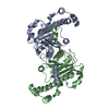 3oteS S: Starting model for refinement |
|---|---|
| Similar structure data |


-Links
 PDBj
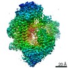
PDBj- Assembly
| Deposited unit | 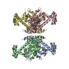
| |||||||||||||||||||||||||||||||||||||||||||||||||||||||||||||||||||||||||||||||||||||||||||||||
|---|---|---|---|---|---|---|---|---|---|---|---|---|---|---|---|---|---|---|---|---|---|---|---|---|---|---|---|---|---|---|---|---|---|---|---|---|---|---|---|---|---|---|---|---|---|---|---|---|---|---|---|---|---|---|---|---|---|---|---|---|---|---|---|---|---|---|---|---|---|---|---|---|---|---|---|---|---|---|---|---|---|---|---|---|---|---|---|---|---|---|---|---|---|---|---|---|
| 1 |
| |||||||||||||||||||||||||||||||||||||||||||||||||||||||||||||||||||||||||||||||||||||||||||||||
| Unit cell |
| |||||||||||||||||||||||||||||||||||||||||||||||||||||||||||||||||||||||||||||||||||||||||||||||
| Noncrystallographic symmetry (NCS) | NCS domain:
NCS domain segments: Ens-ID: 1
|