 Movie
Movie Controller


+ Open data

- Basic information
| Entry | Database: PDB / ID: 7cq4 | ||||||||||||||||||||||||
|---|---|---|---|---|---|---|---|---|---|---|---|---|---|---|---|---|---|---|---|---|---|---|---|---|---|
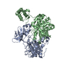
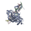
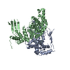
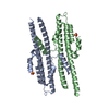
| Title | Crystal structure of Slx1-Slx4 in complex with 5'flap DNA | ||||||||||||||||||||||||
 Components Components |
| ||||||||||||||||||||||||
 Keywords Keywords | HYDROLASE / endonuclease complex / Holliday Junction / SLX1-SLX4 | ||||||||||||||||||||||||
| Function / homology |  Function and homology information Function and homology informationSlx1-Slx4 complex / crossover junction DNA endonuclease activity / 5'-flap endonuclease activity / double-strand break repair via homologous recombination / Hydrolases; Acting on ester bonds / zinc ion binding Similarity search - Function | ||||||||||||||||||||||||
| Biological species |  | ||||||||||||||||||||||||
| Method |  X-RAY DIFFRACTION / SYNCHROTRON / MOLECULAR REPLACEMENT / Resolution: 3.294 Å X-RAY DIFFRACTION / SYNCHROTRON / MOLECULAR REPLACEMENT / Resolution: 3.294 Å | ||||||||||||||||||||||||
 Authors Authors | Xu, X. / Wang, M. / Sun, J. / Li, G. / Yang, N. / Xu, R.M. | ||||||||||||||||||||||||
| Funding support |  China, 7items China, 7items
| ||||||||||||||||||||||||
 Citation Citation | Journal: Nucleic Acids Res. / Year: 2021 Title: Structure specific DNA recognition by the SLX1-SLX4 endonuclease complex. Authors: Xu, X. / Wang, M. / Sun, J. / Yu, Z. / Li, G. / Yang, N. / Xu, R.M. | ||||||||||||||||||||||||
| History |
|
- Structure visualization
| Structure viewer | Molecule: MolmilJmol/JSmol |
|---|
- Downloads & links
-Download
| PDBx/mmCIF format | 7cq4.cif.gz | 133.2 KB | Display | PDBx/mmCIF format |
|---|---|---|---|---|
| PDB format | pdb7cq4.ent.gz | 96.7 KB | Display | PDB format |
| PDBx/mmJSON format | 7cq4.json.gz | Tree view | PDBx/mmJSON format | |
| Others |  Other downloads Other downloads |
-Validation report
| Arichive directory | https://data.pdbj.org/pub/pdb/validation_reports/cq/7cq4ftp://data.pdbj.org/pub/pdb/validation_reports/cq/7cq4 | HTTPS FTP |
|---|
-Related structure data
| Related structure data | 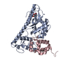 7cq2SC  7cq3C S: Starting model for refinement C: citing same article ( |
|---|---|
| Similar structure data |


-Links
 PDBj
PDBj


- Assembly
| Deposited unit | 
| ||||||||
|---|---|---|---|---|---|---|---|---|---|
| 1 |
| ||||||||
| Unit cell |
|
-Components
-DNA chain , 3 types, 3 molecules HKL
| #1: DNA chain | Mass: 4312.842 Da / Num. of mol.: 1 / Source method: obtained synthetically / Source: (synth.) |
|---|---|
| #2: DNA chain | Mass: 4255.778 Da / Num. of mol.: 1 / Source method: obtained synthetically / Source: (synth.) |
| #3: DNA chain | Mass: 8291.358 Da / Num. of mol.: 1 / Source method: obtained synthetically / Source: (synth.) |
-Protein , 2 types, 2 molecules AB
| #4: Protein | Mass: 35884.621 Da / Num. of mol.: 1 / Mutation: Y17F Source method: isolated from a genetically manipulated source Source: (gene. exp.) Strain: YJM789 / Gene: SLX1, SCY_0436 / Plasmid: pCDF-Duet / Production host:  References: UniProt: A6ZLG6, Hydrolases; Acting on ester bonds |
|---|---|
| #5: Protein | Mass: 17144.518 Da / Num. of mol.: 1 Source method: isolated from a genetically manipulated source Source: (gene. exp.) Gene: SLX4, GI526_G0003928 / Plasmid: pCDF-Duet / Production host: |
-Non-polymers , 2 types, 14 molecules 

| #6: Chemical |  Mass: 65.409 Da / Num. of mol.: 2 / Source method: obtained synthetically / Formula: Zn / Feature type: SUBJECT OF INVESTIGATION Mass: 65.409 Da / Num. of mol.: 2 / Source method: obtained synthetically / Formula: Zn / Feature type: SUBJECT OF INVESTIGATION#7: Water | ChemComp-HOH / | Mass: 18.015 Da / Num. of mol.: 12 / Source method: isolated from a natural source / Formula: H2O |
|---|
-Details
| Has ligand of interest | Y |
|---|
-Experimental details
-Experiment
| Experiment | Method: X-RAY DIFFRACTION / Number of used crystals: 1 |
|---|
- Sample preparation
| Crystal | Density Matthews: 3.66 Å3/Da / Density % sol: 66.41 % |
|---|---|
| Crystal grow | Temperature: 277 K / Method: vapor diffusion / pH: 6 Details: 0.1 M sodium cacodylate, pH 6.0, 12% PEG 1500 and 0.1 M TCEP hydrochloride |
-Data collection
| Diffraction | Mean temperature: 100 K / Serial crystal experiment: N | |||||||||||||||||||||||||||||||||||||||||||||||||||||||||||||||||||||||||||||||||||||||||||||||||||
|---|---|---|---|---|---|---|---|---|---|---|---|---|---|---|---|---|---|---|---|---|---|---|---|---|---|---|---|---|---|---|---|---|---|---|---|---|---|---|---|---|---|---|---|---|---|---|---|---|---|---|---|---|---|---|---|---|---|---|---|---|---|---|---|---|---|---|---|---|---|---|---|---|---|---|---|---|---|---|---|---|---|---|---|---|---|---|---|---|---|---|---|---|---|---|---|---|---|---|---|---|
| Diffraction source | Source: SYNCHROTRON / Site: SSRF / Beamline: BL17U / Wavelength: 0.9791 Å | |||||||||||||||||||||||||||||||||||||||||||||||||||||||||||||||||||||||||||||||||||||||||||||||||||
| Detector | Type: ADSC QUANTUM 315r / Detector: CCD / Date: Dec 13, 2014 | |||||||||||||||||||||||||||||||||||||||||||||||||||||||||||||||||||||||||||||||||||||||||||||||||||
| Radiation | Protocol: SINGLE WAVELENGTH / Monochromatic (M) / Laue (L): M / Scattering type: x-ray | |||||||||||||||||||||||||||||||||||||||||||||||||||||||||||||||||||||||||||||||||||||||||||||||||||
| Radiation wavelength | Wavelength: 0.9791 Å / Relative weight: 1 | |||||||||||||||||||||||||||||||||||||||||||||||||||||||||||||||||||||||||||||||||||||||||||||||||||
| Reflection | Resolution: 3.294→50 Å / Num. obs: 16540 / % possible obs: 99.8 % / Redundancy: 10.9 % / Biso Wilson estimate: 96.37 Å2 / Rmerge(I) obs: 0.122 / Rpim(I) all: 0.039 / Rrim(I) all: 0.128 / Χ2: 1.003 / Net I/σ(I): 7.1 | |||||||||||||||||||||||||||||||||||||||||||||||||||||||||||||||||||||||||||||||||||||||||||||||||||
| Reflection shell | Diffraction-ID: 1
|
- Processing
| Software |
| ||||||||||||||||||||||||||||||||||||||||||
|---|---|---|---|---|---|---|---|---|---|---|---|---|---|---|---|---|---|---|---|---|---|---|---|---|---|---|---|---|---|---|---|---|---|---|---|---|---|---|---|---|---|---|---|
| Refinement | Method to determine structure: MOLECULAR REPLACEMENT Starting model: 7cq2 Resolution: 3.294→48.314 Å / SU ML: 0.52 / Cross valid method: THROUGHOUT / σ(F): 1.34 / Phase error: 27.81 / Stereochemistry target values: ML
| ||||||||||||||||||||||||||||||||||||||||||
| Solvent computation | Shrinkage radii: 0.9 Å / VDW probe radii: 1.11 Å / Solvent model: FLAT BULK SOLVENT MODEL | ||||||||||||||||||||||||||||||||||||||||||
| Displacement parameters | Biso max: 167.84 Å2 / Biso mean: 90.05 Å2 / Biso min: 27.59 Å2 | ||||||||||||||||||||||||||||||||||||||||||
| Refinement step | Cycle: final / Resolution: 3.294→48.314 Å
| ||||||||||||||||||||||||||||||||||||||||||
| Refine LS restraints |
| ||||||||||||||||||||||||||||||||||||||||||
| LS refinement shell | Refine-ID: X-RAY DIFFRACTION / Rfactor Rfree error: 0
|
