 Movie
Movie Controller

[English] 日本語
 Yorodumi
- PDB-7cp0: Crystal Structure of double mutant Y115E Y117E human Secretory Gl... -
+ Open data

- Basic information
| Entry | Database: PDB / ID: 7cp0 | ||||||
|---|---|---|---|---|---|---|---|
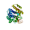
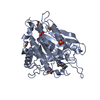
| Title | Crystal Structure of double mutant Y115E Y117E human Secretory Glutaminyl Cyclase | ||||||
 Components Components | Glutaminyl-peptide cyclotransferase | ||||||
 Keywords Keywords | TRANSFERASE / Glutaminyl-peptide cyclotransferase / sQC / Alzheimer's disease / Pyroglutamate | ||||||
| Function / homology |  Function and homology information Function and homology informationpeptidyl-pyroglutamic acid biosynthetic process, using glutaminyl-peptide cyclotransferase / glutaminyl-peptide cyclotransferase / glutaminyl-peptide cyclotransferase activity / protein modification process / specific granule lumen / tertiary granule lumen / ficolin-1-rich granule lumen / Neutrophil degranulation / extracellular exosome / extracellular region / zinc ion binding Similarity search - Function | ||||||
| Biological species |  Homo sapiens (human) Homo sapiens (human) | ||||||
| Method |  X-RAY DIFFRACTION / SYNCHROTRON / MOLECULAR REPLACEMENT / Resolution: 1.7 Å X-RAY DIFFRACTION / SYNCHROTRON / MOLECULAR REPLACEMENT / Resolution: 1.7 Å | ||||||
 Authors Authors | Dileep, K.V. / Ihara, K. / Sakai, N. / Shirozu, M. / Zhang, K.Y.J. | ||||||
| Funding support |  Japan, 1items Japan, 1items
| ||||||
 Citation Citation | Journal: Int.J.Biol.Macromol. / Year: 2020 Title: Piperidine-4-carboxamide as a new scaffold for designing secretory glutaminyl cyclase inhibitors. Authors: Dileep, K.V. / Sakai, N. / Ihara, K. / Kato-Murayama, M. / Nakata, A. / Ito, A. / Sivaraman, D.M. / Shin, J.W. / Yoshida, M. / Shirouzu, M. / Zhang, K.Y.J. | ||||||
| History |
|
- Structure visualization
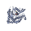
| Structure viewer | Molecule: MolmilJmol/JSmol |
|---|
- Downloads & links
-Download
| PDBx/mmCIF format | 7cp0.cif.gz | 220 KB | Display | PDBx/mmCIF format |
|---|---|---|---|---|
| PDB format | pdb7cp0.ent.gz | 175.1 KB | Display | PDB format |
| PDBx/mmJSON format | 7cp0.json.gz | Tree view | PDBx/mmJSON format | |
| Others |  Other downloads Other downloads |
-Validation report
| Arichive directory | https://data.pdbj.org/pub/pdb/validation_reports/cp/7cp0ftp://data.pdbj.org/pub/pdb/validation_reports/cp/7cp0 | HTTPS FTP |
|---|
-Related structure data
| Related structure data |  7cozC  4ywyS S: Starting model for refinement C: citing same article ( |
|---|---|
| Similar structure data |


-Links
 PDBj
PDBj
- Assembly
| Deposited unit | 
| |||||||||
|---|---|---|---|---|---|---|---|---|---|---|
| 1 | 
| |||||||||
| 2 | 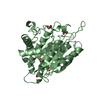
| |||||||||
| 3 | 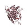
| |||||||||
| Unit cell |
| |||||||||
| Components on special symmetry positions |
|
-Components
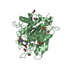
| #1: Protein | Mass: 37331.121 Da / Num. of mol.: 3 / Mutation: Y115E, Y117E Source method: isolated from a genetically manipulated source Source: (gene. exp.) Homo sapiens (human) / Gene: QPCTProduction host:  References: UniProt: Q16769, glutaminyl-peptide cyclotransferase #2: Chemical |   Mass: 65.409 Da / Num. of mol.: 3 / Source method: obtained synthetically / Formula: Zn Mass: 65.409 Da / Num. of mol.: 3 / Source method: obtained synthetically / Formula: Zn#3: Chemical |   Mass: 96.063 Da / Num. of mol.: 3 / Source method: obtained synthetically / Formula: SO4 Mass: 96.063 Da / Num. of mol.: 3 / Source method: obtained synthetically / Formula: SO4#4: Chemical | ChemComp-EDO /   Mass: 62.068 Da / Num. of mol.: 21 / Source method: obtained synthetically / Formula: C2H6O2 Mass: 62.068 Da / Num. of mol.: 21 / Source method: obtained synthetically / Formula: C2H6O2#5: Water | ChemComp-HOH / |  Mass: 18.015 Da / Num. of mol.: 648 / Source method: isolated from a natural source / Formula: H2O Mass: 18.015 Da / Num. of mol.: 648 / Source method: isolated from a natural source / Formula: H2OHas ligand of interest | N | |
|---|
-Experimental details
-Experiment
| Experiment | Method: X-RAY DIFFRACTION / Number of used crystals: 1 |
|---|
- Sample preparation
| Crystal | Density Matthews: 2.75 Å3/Da / Density % sol: 55.23 % / Description: Hexagonal plate like crystals |
|---|---|
| Crystal grow | Temperature: 277.15 K / Method: vapor diffusion, sitting drop / pH: 6 Details: 100 mM MES buffer pH 6.5, 46-67 mM NaOH, 100 mM ammonium sulfate PH range: 6.0-6.5 |
-Data collection
| Diffraction | Mean temperature: 100 K / Serial crystal experiment: N | ||||||||||||||||||||||||
|---|---|---|---|---|---|---|---|---|---|---|---|---|---|---|---|---|---|---|---|---|---|---|---|---|---|
| Diffraction source | Source: SYNCHROTRON / Site: SPring-8 / Beamline: BL26B2 / Wavelength: 1 Å | ||||||||||||||||||||||||
| Detector | Type: RAYONIX MX-225 / Detector: CCD / Date: Jul 21, 2017 | ||||||||||||||||||||||||
| Radiation | Protocol: SINGLE WAVELENGTH / Monochromatic (M) / Laue (L): M / Scattering type: x-ray | ||||||||||||||||||||||||
| Radiation wavelength | Wavelength: 1 Å / Relative weight: 1 | ||||||||||||||||||||||||
| Reflection | Resolution: 1.7→43.05 Å / Num. obs: 131782 / % possible obs: 99.8 % / Redundancy: 3.8 % / CC1/2: 0.996 / Rmerge(I) obs: 0.09 / Net I/σ(I): 10.6 / Num. measured all: 494444 / Scaling rejects: 151 | ||||||||||||||||||||||||
| Reflection shell | Diffraction-ID: 1
|
- Processing
| Software |
| ||||||||||||||||||||||||
|---|---|---|---|---|---|---|---|---|---|---|---|---|---|---|---|---|---|---|---|---|---|---|---|---|---|
| Refinement | Method to determine structure: MOLECULAR REPLACEMENT Starting model: 4YWY Resolution: 1.7→43.05 Å / Cor.coef. Fo:Fc: 0.941 / Cor.coef. Fo:Fc free: 0.931 / SU B: 0.001 / SU ML: 0 / Cross valid method: THROUGHOUT / σ(F): 0 / ESU R: 0.093 / ESU R Free: 0.101 / Stereochemistry target values: MAXIMUM LIKELIHOOD Details: HYDROGENS HAVE BEEN ADDED IN THE RIDING POSITIONS U VALUES : REFINED INDIVIDUALLY
| ||||||||||||||||||||||||
| Solvent computation | Ion probe radii: 0.8 Å / Shrinkage radii: 0.8 Å / VDW probe radii: 1.2 Å / Solvent model: MASK | ||||||||||||||||||||||||
| Displacement parameters | Biso max: 61.93 Å2 / Biso mean: 17.651 Å2 / Biso min: 6.83 Å2
| ||||||||||||||||||||||||
| Refinement step | Cycle: final / Resolution: 1.7→43.05 Å
| ||||||||||||||||||||||||
| LS refinement shell | Resolution: 1.7→1.744 Å / Rfactor Rfree error: 0 / Total num. of bins used: 20
|
