 Movie
Movie Controller

[English] 日本語
 Yorodumi
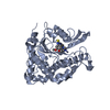
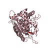
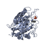
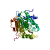
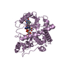
- PDB-6gbx: Crystal structure of human glutaminyl cyclase variant Y115E-Y117E... -
+ Open data

- Basic information
| Entry | Database: PDB / ID: 6gbx | ||||||
|---|---|---|---|---|---|---|---|
| Title | Crystal structure of human glutaminyl cyclase variant Y115E-Y117E in complex with SEN177 | ||||||
 Components Components | Glutaminyl-peptide cyclotransferase | ||||||
 Keywords Keywords | TRANSFERASE / Inhibitor / Cyclotransferase / zinc enzyme / Alzheimer | ||||||
| Function / homology |  Function and homology information Function and homology informationpeptidyl-pyroglutamic acid biosynthetic process, using glutaminyl-peptide cyclotransferase / glutaminyl-peptide cyclotransferase / glutaminyl-peptide cyclotransferase activity / protein modification process / specific granule lumen / tertiary granule lumen / ficolin-1-rich granule lumen / Neutrophil degranulation / extracellular exosome / extracellular region / zinc ion binding Similarity search - Function | ||||||
| Biological species |  Homo sapiens (human) Homo sapiens (human) | ||||||
| Method |  X-RAY DIFFRACTION / SYNCHROTRON / MOLECULAR REPLACEMENT / Resolution: 1.72 Å X-RAY DIFFRACTION / SYNCHROTRON / MOLECULAR REPLACEMENT / Resolution: 1.72 Å | ||||||
 Authors Authors | Pozzi, C. / Di Pisa, F. / Benvenuti, M. / Mangani, S. | ||||||
 Citation Citation | Journal: J. Biol. Inorg. Chem. / Year: 2018 Title: The structure of the human glutaminyl cyclase-SEN177 complex indicates routes for developing new potent inhibitors as possible agents for the treatment of neurological disorders. Authors: Pozzi, C. / Di Pisa, F. / Benvenuti, M. / Mangani, S. | ||||||
| History |
|
- Structure visualization
| Structure viewer | Molecule: MolmilJmol/JSmol |
|---|
- Downloads & links
-Download
| PDBx/mmCIF format | 6gbx.cif.gz | 226.4 KB | Display | PDBx/mmCIF format |
|---|---|---|---|---|
| PDB format | pdb6gbx.ent.gz | 180.1 KB | Display | PDB format |
| PDBx/mmJSON format | 6gbx.json.gz | Tree view | PDBx/mmJSON format | |
| Others |  Other downloads Other downloads |
-Validation report
| Arichive directory | https://data.pdbj.org/pub/pdb/validation_reports/gb/6gbxftp://data.pdbj.org/pub/pdb/validation_reports/gb/6gbx | HTTPS FTP |
|---|
-Related structure data
| Related structure data |  4yu9S S: Starting model for refinement |
|---|---|
| Similar structure data |


-Links
 PDBj
PDBj
- Assembly
| Deposited unit | 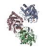
| |||||||||
|---|---|---|---|---|---|---|---|---|---|---|
| 1 | 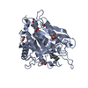
| |||||||||
| 2 | 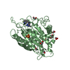
| |||||||||
| 3 | 
| |||||||||
| Unit cell |
| |||||||||
| Components on special symmetry positions |
|
-Components
-Protein , 1 types, 3 molecules ABC
| #1: Protein | Mass: 37489.277 Da / Num. of mol.: 3 / Mutation: Y115E; Y117E Source method: isolated from a genetically manipulated source Source: (gene. exp.) Homo sapiens (human) / Gene: QPCT / Production host:  References: UniProt: Q16769, glutaminyl-peptide cyclotransferase |
|---|
-Non-polymers , 5 types, 726 molecules 


| #2: Chemical |  Mass: 65.409 Da / Num. of mol.: 3 / Source method: obtained synthetically / Formula: Zn Mass: 65.409 Da / Num. of mol.: 3 / Source method: obtained synthetically / Formula: Zn#3: Chemical |  Mass: 96.063 Da / Num. of mol.: 3 / Source method: obtained synthetically / Formula: SO4 Mass: 96.063 Da / Num. of mol.: 3 / Source method: obtained synthetically / Formula: SO4#4: Chemical |  Mass: 338.382 Da / Num. of mol.: 3 / Source method: obtained synthetically / Formula: C18H19FN6 / Feature type: SUBJECT OF INVESTIGATION Mass: 338.382 Da / Num. of mol.: 3 / Source method: obtained synthetically / Formula: C18H19FN6 / Feature type: SUBJECT OF INVESTIGATION#5: Chemical | ChemComp-EDO /  Mass: 62.068 Da / Num. of mol.: 20 / Source method: obtained synthetically / Formula: C2H6O2 Mass: 62.068 Da / Num. of mol.: 20 / Source method: obtained synthetically / Formula: C2H6O2#6: Water | ChemComp-HOH / | Mass: 18.015 Da / Num. of mol.: 697 / Source method: isolated from a natural source / Formula: H2O |
|---|
-Experimental details
-Experiment
| Experiment | Method: X-RAY DIFFRACTION / Number of used crystals: 1 |
|---|
- Sample preparation
| Crystal | Density Matthews: 2.44 Å3/Da / Density % sol: 49.6 % |
|---|---|
| Crystal grow | Temperature: 277.15 K / Method: vapor diffusion, hanging drop / pH: 6.5 Details: 0.1 M MES, 0.4 M ammonium sulfate, 0.07 M sodium chloride |
-Data collection
| Diffraction | Mean temperature: 100 K |
|---|---|
| Diffraction source | Source: SYNCHROTRON / Site: Diamond  / Beamline: I04-1 / Wavelength: 0.9795 Å / Beamline: I04-1 / Wavelength: 0.9795 Å |
| Detector | Type: DECTRIS PILATUS 2M / Detector: PIXEL / Date: Sep 28, 2013 |
| Radiation | Protocol: SINGLE WAVELENGTH / Monochromatic (M) / Laue (L): M / Scattering type: x-ray |
| Radiation wavelength | Wavelength: 0.9795 Å / Relative weight: 1 |
| Reflection | Resolution: 1.72→95.23 Å / Num. obs: 126884 / % possible obs: 99.8 % / Redundancy: 3.4 % / Biso Wilson estimate: 9.28 Å2 / CC1/2: 0.985 / Rrim(I) all: 0.13 / Net I/σ(I): 5.3 |
| Reflection shell | Resolution: 1.72→1.75 Å / Redundancy: 2.9 % / Mean I/σ(I) obs: 1.7 / Num. unique obs: 6232 / CC1/2: 0.567 / Rrim(I) all: 0.75 / % possible all: 99.4 |
- Processing
| Software |
| ||||||||||||||||||||||||||||||||||||||||||||||||||||||||||||||||||||||||||||||||||||||||||||||||||||||||||||||||||||||||||||||||||||||||||||||||||||||||||||||||||||||||||||||||||||||
|---|---|---|---|---|---|---|---|---|---|---|---|---|---|---|---|---|---|---|---|---|---|---|---|---|---|---|---|---|---|---|---|---|---|---|---|---|---|---|---|---|---|---|---|---|---|---|---|---|---|---|---|---|---|---|---|---|---|---|---|---|---|---|---|---|---|---|---|---|---|---|---|---|---|---|---|---|---|---|---|---|---|---|---|---|---|---|---|---|---|---|---|---|---|---|---|---|---|---|---|---|---|---|---|---|---|---|---|---|---|---|---|---|---|---|---|---|---|---|---|---|---|---|---|---|---|---|---|---|---|---|---|---|---|---|---|---|---|---|---|---|---|---|---|---|---|---|---|---|---|---|---|---|---|---|---|---|---|---|---|---|---|---|---|---|---|---|---|---|---|---|---|---|---|---|---|---|---|---|---|---|---|---|---|
| Refinement | Method to determine structure: MOLECULAR REPLACEMENT Starting model: 4yu9 Resolution: 1.72→95.23 Å / Cor.coef. Fo:Fc: 0.96 / Cor.coef. Fo:Fc free: 0.95 / SU B: 2.126 / SU ML: 0.066 / Cross valid method: THROUGHOUT / ESU R: 0.089 / ESU R Free: 0.087 / Details: HYDROGENS HAVE BEEN ADDED IN THE RIDING POSITIONS
| ||||||||||||||||||||||||||||||||||||||||||||||||||||||||||||||||||||||||||||||||||||||||||||||||||||||||||||||||||||||||||||||||||||||||||||||||||||||||||||||||||||||||||||||||||||||
| Solvent computation | Ion probe radii: 0.8 Å / Shrinkage radii: 0.8 Å / VDW probe radii: 1.2 Å | ||||||||||||||||||||||||||||||||||||||||||||||||||||||||||||||||||||||||||||||||||||||||||||||||||||||||||||||||||||||||||||||||||||||||||||||||||||||||||||||||||||||||||||||||||||||
| Displacement parameters | Biso mean: 15.516 Å2 | ||||||||||||||||||||||||||||||||||||||||||||||||||||||||||||||||||||||||||||||||||||||||||||||||||||||||||||||||||||||||||||||||||||||||||||||||||||||||||||||||||||||||||||||||||||||
| Refinement step | Cycle: 1 / Resolution: 1.72→95.23 Å
| ||||||||||||||||||||||||||||||||||||||||||||||||||||||||||||||||||||||||||||||||||||||||||||||||||||||||||||||||||||||||||||||||||||||||||||||||||||||||||||||||||||||||||||||||||||||
| Refine LS restraints |
|