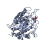
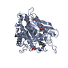
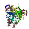
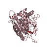
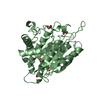
[English] 日本語
Yorodumi
- PDB-4ywy: Crystal Structure of double mutant Y115E Y117E human Glutaminyl C... -

+
Open data


ID or keywords:

Loading...

-
Basic information

Entry
Database: PDB / ID: 4ywy
TitleCrystal Structure of double mutant Y115E Y117E human Glutaminyl Cyclase in complex with inhibitor PBD-150
ComponentsGlutaminyl-peptide cyclotransferase
KeywordsTRANSFERASE / Alzheimer Disease / Cyclotransferase Soluble Variant / PBD-150 inhibitor
Function / homology
Function and homology information


peptidyl-pyroglutamic acid biosynthetic process, using glutaminyl-peptide cyclotransferase / glutaminyl-peptide cyclotransferase / glutaminyl-peptide cyclotransferase activity / protein modification process / specific granule lumen / tertiary granule lumen / ficolin-1-rich granule lumen / Neutrophil degranulation / extracellular exosome / zinc ion binding / extracellular region
Similarity search - Function
M28 Zn-Peptidase Glutaminyl Cyclase / Glutaminyl-peptide cyclotransferase-like / Peptidase M28 / Peptidase family M28 / Zn peptidases / Aminopeptidase / 3-Layer(aba) Sandwich / Alpha Beta
Similarity search - Domain/homology
Chem-PBD / Glutaminyl-peptide cyclotransferase
Similarity search - Component
Biological speciesHomo sapiens (human)
MethodX-RAY DIFFRACTION / SYNCHROTRON / MOLECULAR REPLACEMENT / Resolution: 1.95 Å
AuthorsDi Pisa, F. / Pozzi, C. / Benvenuti, M. / Mangani, S.
CitationJournal: Acta Crystallogr.,Sect.F / Year: 2015
Title: The soluble Y115E-Y117E variant of human glutaminyl cyclase is a valid target for X-ray and NMR screening of inhibitors against Alzheimer disease.
Authors: DiPisa, F. / Pozzi, C. / Benvenuti, M. / Andreini, M. / Marconi, G. / Mangani, S.
History
DepositionMar 21, 2015Deposition site: RCSB / Processing site: PDBE
Revision 1.0Aug 5, 2015Provider: repository / Type: Initial release
Revision 1.1Aug 12, 2015Group: Database references
Revision 1.2Aug 19, 2015Group: Database references
Revision 1.3Jan 10, 2024Group: Data collection / Database references ...Data collection / Database references / Derived calculations / Refinement description
Category: chem_comp_atom / chem_comp_bond ...chem_comp_atom / chem_comp_bond / database_2 / pdbx_initial_refinement_model / struct_conn
Item: _database_2.pdbx_DOI / _database_2.pdbx_database_accession ..._database_2.pdbx_DOI / _database_2.pdbx_database_accession / _struct_conn.pdbx_dist_value / _struct_conn.ptnr1_auth_asym_id / _struct_conn.ptnr1_auth_comp_id / _struct_conn.ptnr1_auth_seq_id / _struct_conn.ptnr1_label_asym_id / _struct_conn.ptnr1_label_atom_id / _struct_conn.ptnr1_label_comp_id / _struct_conn.ptnr1_label_seq_id / _struct_conn.ptnr2_auth_asym_id / _struct_conn.ptnr2_auth_comp_id / _struct_conn.ptnr2_auth_seq_id / _struct_conn.ptnr2_label_asym_id / _struct_conn.ptnr2_label_atom_id / _struct_conn.ptnr2_label_comp_id

-
Structure visualization

Structure viewerMolecule:
MolmilJmol/JSmol

Downloads & links

-
Assembly

Deposited unit
A: Glutaminyl-peptide cyclotransferase
B: Glutaminyl-peptide cyclotransferase
C: Glutaminyl-peptide cyclotransferase
hetero molecules


Theoretical massNumber of molelcules
Total (without water)125,48335
Polymers122,5693
Non-polymers2,91532
Water23,4381301
1
A: Glutaminyl-peptide cyclotransferase
hetero molecules


Theoretical massNumber of molelcules
Total (without water)41,80812
Polymers40,8561
Non-polymers95211
Water181
TypeNameSymmetry operationNumber
identity operation1_555x,y,z1
2
B: Glutaminyl-peptide cyclotransferase
hetero molecules


Theoretical massNumber of molelcules
Total (without water)41,86912
Polymers40,8561
Non-polymers1,01211
Water181
TypeNameSymmetry operationNumber
identity operation1_555x,y,z1
3
C: Glutaminyl-peptide cyclotransferase
hetero molecules


Theoretical massNumber of molelcules
Total (without water)41,80711
Polymers40,8561
Non-polymers95010
Water181
TypeNameSymmetry operationNumber
identity operation1_555x,y,z1
Unit cell
Length a, b, c (Å)86.429, 149.540, 96.210
Angle α, β, γ (deg.)90.00, 96.82, 90.00
Int Tables number5
Space group name H-MC121
Components on special symmetry positions
IDModelComponents
11C-819-

HOH

21C-852-

HOH

-
Components

-
Protein , 1 types, 3 molecules ABC

#1: Protein Glutaminyl-peptide cyclotransferase / / Glutaminyl cyclase / sQC / Glutaminyl-tRNA cyclotransferase / Glutamyl cyclase / EC


Mass: 40856.285 Da / Num. of mol.: 3
Source method: isolated from a genetically manipulated source
Source: (gene. exp.) Homo sapiens (human) / Gene: QPCT / Plasmid: pQE80L / Production host: Escherichia coli (E. coli)
References: UniProt: Q16769, glutaminyl-peptide cyclotransferase

-
Non-polymers , 6 types, 1333 molecules

#2: Chemical ChemComp-ZN / ZINC ION


Mass: 65.409 Da / Num. of mol.: 3 / Source method: obtained synthetically / Formula: Zn
#3: Chemical ChemComp-PBD / 1-(3,4-dimethoxyphenyl)-3-[3-(1H-imidazol-1-yl)propyl]thiourea


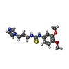
Mass: 320.410 Da / Num. of mol.: 3 / Source method: obtained synthetically / Formula: C15H20N4O2S
#4: Chemical
ChemComp-EDO / 1,2-ETHANEDIOL / ETHYLENE GLYCOL / Ethylene glycol


Mass: 62.068 Da / Num. of mol.: 20 / Source method: obtained synthetically / Formula: C2H6O2
#5: Chemical
ChemComp-SO4 / SULFATE ION / Sulfate


Mass: 96.063 Da / Num. of mol.: 5 / Source method: obtained synthetically / Formula: SO4
#6: Chemical ChemComp-CL / CHLORIDE ION / Chloride


Mass: 35.453 Da / Num. of mol.: 1 / Source method: obtained synthetically / Formula: Cl
#7: Water ChemComp-HOH / water / Water


Mass: 18.015 Da / Num. of mol.: 1301 / Source method: isolated from a natural source / Formula: H2O

-
Experimental details

-
Experiment

ExperimentMethod: X-RAY DIFFRACTION

-
Sample preparation

CrystalDensity Matthews: 2.52 Å3/Da / Density % sol: 51.16 %
Crystal growTemperature: 281 K / Method: vapor diffusion, sitting drop / pH: 6.5 / Details: 0.1 M MES buffer pH 6.5, 1.6 M ammonium sulfate

-
Data collection

DiffractionMean temperature: 100 K
Diffraction sourceSource: SYNCHROTRON / Site: Diamond / Beamline: I04-1 / Wavelength: 0.979 Å
DetectorType: DECTRIS PILATUS 2M / Detector: PIXEL / Date: Oct 18, 2014
RadiationProtocol: SINGLE WAVELENGTH / Monochromatic (M) / Laue (L): M / Scattering type: x-ray
Radiation wavelengthWavelength: 0.979 Å / Relative weight: 1
ReflectionResolution: 1.95→32.99 Å / Num. all: 257040 / Num. obs: 87147 / % possible obs: 99 % / Redundancy: 2.9 % / Net I/σ(I): 6

-
Processing

Software
NameVersionClassification
REFMAC5.8.0073refinement
MOSFLMdata reduction
SCALAdata scaling
MOLREPphasing
RefinementMethod to determine structure: MOLECULAR REPLACEMENT
Starting model: 2AFM
Resolution: 1.95→32.99 Å / Cor.coef. Fo:Fc: 0.946 / Cor.coef. Fo:Fc free: 0.915 / SU B: 3.723 / SU ML: 0.104 / Cross valid method: THROUGHOUT / ESU R: 0.147 / ESU R Free: 0.141 / Stereochemistry target values: MAXIMUM LIKELIHOOD / Details: HYDROGENS HAVE BEEN ADDED IN THE RIDING POSITIONS
RfactorNum. reflection% reflectionSelection details
Rfree0.21101 4184 4.8 %RANDOM
Rwork0.16408 ---
obs0.1663 82798 98.77 %-
Solvent computationIon probe radii: 0.8 Å / Shrinkage radii: 0.8 Å / VDW probe radii: 1.2 Å / Solvent model: MASK
Displacement parametersBiso mean: 13.098 Å2
Baniso -1Baniso -2Baniso -3
1-0 Å2-0 Å20 Å2
2---0 Å2-0 Å2
3----0 Å2
Refinement stepCycle: 1 / Resolution: 1.95→32.99 Å
ProteinNucleic acidLigandSolventTotal
Num. atoms7752 0 175 1301 9228
Refine LS restraints
Refine-IDTypeDev idealDev ideal targetNumber
X-RAY DIFFRACTIONr_bond_refined_d0.0180.0198182
X-RAY DIFFRACTIONr_bond_other_d0.0030.027621
X-RAY DIFFRACTIONr_angle_refined_deg1.9121.9711106
X-RAY DIFFRACTIONr_angle_other_deg0.9843.00317508
X-RAY DIFFRACTIONr_dihedral_angle_1_deg6.6755993
X-RAY DIFFRACTIONr_dihedral_angle_2_deg34.30523.874382
X-RAY DIFFRACTIONr_dihedral_angle_3_deg12.446151273
X-RAY DIFFRACTIONr_dihedral_angle_4_deg19.2881548
X-RAY DIFFRACTIONr_chiral_restr0.110.21204
X-RAY DIFFRACTIONr_gen_planes_refined0.010.0219193
X-RAY DIFFRACTIONr_gen_planes_other0.0010.021932
X-RAY DIFFRACTIONr_nbd_refined
X-RAY DIFFRACTIONr_nbd_other
X-RAY DIFFRACTIONr_nbtor_refined
X-RAY DIFFRACTIONr_nbtor_other
X-RAY DIFFRACTIONr_xyhbond_nbd_refined
X-RAY DIFFRACTIONr_xyhbond_nbd_other
X-RAY DIFFRACTIONr_metal_ion_refined
X-RAY DIFFRACTIONr_metal_ion_other
X-RAY DIFFRACTIONr_symmetry_vdw_refined
X-RAY DIFFRACTIONr_symmetry_vdw_other
X-RAY DIFFRACTIONr_symmetry_hbond_refined
X-RAY DIFFRACTIONr_symmetry_hbond_other
X-RAY DIFFRACTIONr_symmetry_metal_ion_refined
X-RAY DIFFRACTIONr_symmetry_metal_ion_other
X-RAY DIFFRACTIONr_mcbond_it1.3421.1463930
X-RAY DIFFRACTIONr_mcbond_other1.3421.1453929
X-RAY DIFFRACTIONr_mcangle_it2.2471.7064907
X-RAY DIFFRACTIONr_mcangle_other2.2471.7064908
X-RAY DIFFRACTIONr_scbond_it1.7841.294252
X-RAY DIFFRACTIONr_scbond_other1.7711.2854248
X-RAY DIFFRACTIONr_scangle_it
X-RAY DIFFRACTIONr_scangle_other2.8041.8596184
X-RAY DIFFRACTIONr_long_range_B_refined6.00610.65610585
X-RAY DIFFRACTIONr_long_range_B_other5.6139.8869918
X-RAY DIFFRACTIONr_rigid_bond_restr
X-RAY DIFFRACTIONr_sphericity_free
X-RAY DIFFRACTIONr_sphericity_bonded
LS refinement shellResolution: 1.95→2.001 Å / Total num. of bins used: 20
RfactorNum. reflection% reflection
Rfree0.259 330 -
Rwork0.214 6094 -
obs--98.79 %

+
About Yorodumi

-
News

-
Feb 9, 2022. New format data for meta-information of EMDB entries

New format data for meta-information of EMDB entries

  • Version 3 of the EMDB header file is now the official format.
  • The previous official version 1.9 will be removed from the archive.

Related info.:EMDB header

External links:wwPDB to switch to version 3 of the EMDB data model

-
Aug 12, 2020. Covid-19 info

Covid-19 info

URL: https://pdbj.org/emnavi/covid19.php

New page: Covid-19 featured information page in EM Navigator.

Related info.:Covid-19 info / Mar 5, 2020. Novel coronavirus structure data

+
Mar 5, 2020. Novel coronavirus structure data

Novel coronavirus structure data

Related info.:Yorodumi Speices / Aug 12, 2020. Covid-19 info

External links:COVID-19 featured content - PDBj / Molecule of the Month (242):Coronavirus Proteases

+
Jan 31, 2019. EMDB accession codes are about to change! (news from PDBe EMDB page)

EMDB accession codes are about to change! (news from PDBe EMDB page)

  • The allocation of 4 digits for EMDB accession codes will soon come to an end. Whilst these codes will remain in use, new EMDB accession codes will include an additional digit and will expand incrementally as the available range of codes is exhausted. The current 4-digit format prefixed with “EMD-” (i.e. EMD-XXXX) will advance to a 5-digit format (i.e. EMD-XXXXX), and so on. It is currently estimated that the 4-digit codes will be depleted around Spring 2019, at which point the 5-digit format will come into force.
  • The EM Navigator/Yorodumi systems omit the EMD- prefix.

Related info.:Q: What is EMD? / ID/Accession-code notation in Yorodumi/EM Navigator

External links:EMDB Accession Codes are Changing Soon! / Contact to PDBj

+
Jul 12, 2017. Major update of PDB

Major update of PDB

  • wwPDB released updated PDB data conforming to the new PDBx/mmCIF dictionary.
  • This is a major update changing the version number from 4 to 5, and with Remediation, in which all the entries are updated.
  • In this update, many items about electron microscopy experimental information are reorganized (e.g. em_software).
  • Now, EM Navigator and Yorodumi are based on the updated data.

External links:wwPDB Remediation / Enriched Model Files Conforming to OneDep Data Standards Now Available in the PDB FTP Archive

-
Yorodumi

Thousand views of thousand structures

  • Yorodumi is a browser for structure data from EMDB, PDB, SASBDB, etc.
  • This page is also the successor to EM Navigator detail page, and also detail information page/front-end page for Omokage search.
  • The word "yorodu" (or yorozu) is an old Japanese word meaning "ten thousand". "mi" (miru) is to see.

Related info.:EMDB / PDB / SASBDB / Comparison of 3 databanks / Yorodumi Search / Aug 31, 2016. New EM Navigator & Yorodumi / Yorodumi Papers / Jmol/JSmol / Function and homology information / Changes in new EM Navigator and Yorodumi

Read more