 Movie
Movie Controller


+ Open data

- Basic information
| Entry | Database: PDB / ID: 2afm | ||||||
|---|---|---|---|---|---|---|---|
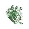
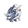
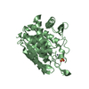
| Title | Crystal structure of human glutaminyl cyclase at pH 6.5 | ||||||
 Components Components | Glutaminyl-peptide cyclotransferase | ||||||
 Keywords Keywords | TRANSFERASE / alpha-beta protein / metalloprotein | ||||||
| Function / homology |  Function and homology information Function and homology informationpeptidyl-pyroglutamic acid biosynthetic process, using glutaminyl-peptide cyclotransferase / glutaminyl-peptide cyclotransferase / glutaminyl-peptide cyclotransferase activity / protein modification process / specific granule lumen / tertiary granule lumen / ficolin-1-rich granule lumen / Neutrophil degranulation / extracellular exosome / extracellular region / zinc ion binding Similarity search - Function | ||||||
| Biological species |  Homo sapiens (human) Homo sapiens (human) | ||||||
| Method |  X-RAY DIFFRACTION / SYNCHROTRON / MAD / Resolution: 1.66 Å X-RAY DIFFRACTION / SYNCHROTRON / MAD / Resolution: 1.66 Å | ||||||
 Authors Authors | Huang, K.F. / Liu, Y.L. / Cheng, W.J. / Ko, T.P. / Wang, A.H.J. | ||||||
 Citation Citation | Journal: Proc.Natl.Acad.Sci.Usa / Year: 2005 Title: Crystal structures of human glutaminyl cyclase, an enzyme responsible for protein N-terminal pyroglutamate formation Authors: Huang, K.F. / Liu, Y.L. / Cheng, W.J. / Ko, T.P. / Wang, A.H. | ||||||
| History |
|
- Structure visualization
| Structure viewer | Molecule: MolmilJmol/JSmol |
|---|
- Downloads & links
-Download
| PDBx/mmCIF format | 2afm.cif.gz | 153.8 KB | Display | PDBx/mmCIF format |
|---|---|---|---|---|
| PDB format | pdb2afm.ent.gz | 120.5 KB | Display | PDB format |
| PDBx/mmJSON format | 2afm.json.gz | Tree view | PDBx/mmJSON format | |
| Others |  Other downloads Other downloads |
-Validation report
| Arichive directory | https://data.pdbj.org/pub/pdb/validation_reports/af/2afmftp://data.pdbj.org/pub/pdb/validation_reports/af/2afm | HTTPS FTP |
|---|
-Related structure data
| Related structure data |  2afoC  2afsC 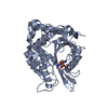 2afuC  2afwC  2afxC 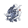 2afzC C: citing same article ( |
|---|---|
| Similar structure data |


-Links
 PDBj
PDBj
- Assembly
| Deposited unit | 
| ||||||||||||||||||||||||
|---|---|---|---|---|---|---|---|---|---|---|---|---|---|---|---|---|---|---|---|---|---|---|---|---|---|
| 1 | 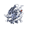
| ||||||||||||||||||||||||
| 2 | 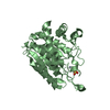
| ||||||||||||||||||||||||
| 3 | 
| ||||||||||||||||||||||||
| Unit cell |
| ||||||||||||||||||||||||
| Components on special symmetry positions |
|
-Components
| #1: Protein | Mass: 37557.398 Da / Num. of mol.: 2 / Fragment: residues 33-361 Source method: isolated from a genetically manipulated source Source: (gene. exp.) Homo sapiens (human) / Gene: QPCT / Plasmid: pET32a / Production host:  References: UniProt: Q16769, glutaminyl-peptide cyclotransferase #2: Chemical |   Mass: 65.409 Da / Num. of mol.: 2 / Source method: obtained synthetically / Formula: Zn Mass: 65.409 Da / Num. of mol.: 2 / Source method: obtained synthetically / Formula: Zn#3: Chemical |   Mass: 96.063 Da / Num. of mol.: 2 / Source method: obtained synthetically / Formula: SO4 Mass: 96.063 Da / Num. of mol.: 2 / Source method: obtained synthetically / Formula: SO4#4: Water | ChemComp-HOH / |  Mass: 18.015 Da / Num. of mol.: 602 / Source method: isolated from a natural source / Formula: H2O Mass: 18.015 Da / Num. of mol.: 602 / Source method: isolated from a natural source / Formula: H2O |
|---|
-Experimental details
-Experiment
| Experiment | Method: X-RAY DIFFRACTION / Number of used crystals: 1 |
|---|
- Sample preparation
| Crystal | Density Matthews: 3.02 Å3/Da / Density % sol: 58 % |
|---|---|
| Crystal grow | Temperature: 298 K / Method: vapor diffusion, hanging drop / pH: 6.5 Details: ammonium sulfate, dioxane, MES, pH 6.5, VAPOR DIFFUSION, HANGING DROP, temperature 298.0K |
-Data collection
| Diffraction | Mean temperature: 123 K | ||||||||||||
|---|---|---|---|---|---|---|---|---|---|---|---|---|---|
| Diffraction source | Source: SYNCHROTRON / Site: Photon Factory  / Beamline: BL-5A / Wavelength: 0.9792, 0.9794, 0.9750 / Beamline: BL-5A / Wavelength: 0.9792, 0.9794, 0.9750 | ||||||||||||
| Detector | Type: ADSC QUANTUM 4 / Detector: CCD / Date: Jun 13, 2004 | ||||||||||||
| Radiation | Monochromator: GRAPHITE / Protocol: MAD / Monochromatic (M) / Laue (L): M / Scattering type: x-ray | ||||||||||||
| Radiation wavelength |
| ||||||||||||
| Reflection | Resolution: 1.66→50 Å / Num. all: 107174 / Num. obs: 106852 / % possible obs: 99.7 % / Observed criterion σ(F): 2 / Observed criterion σ(I): 2 / Redundancy: 5.7 % / Rmerge(I) obs: 0.047 / Net I/σ(I): 36.9 | ||||||||||||
| Reflection shell | Resolution: 1.66→1.72 Å / Redundancy: 5.1 % / Rmerge(I) obs: 0.49 / Mean I/σ(I) obs: 3.5 / % possible all: 100 |
- Processing
| Software |
| ||||||||||||||||||||
|---|---|---|---|---|---|---|---|---|---|---|---|---|---|---|---|---|---|---|---|---|---|
| Refinement | Method to determine structure: MAD / Resolution: 1.66→50 Å / σ(F): 0 / Stereochemistry target values: Engh & Huber
| ||||||||||||||||||||
| Refinement step | Cycle: LAST / Resolution: 1.66→50 Å
| ||||||||||||||||||||
| Refine LS restraints |
|
