 Movie
Movie Controller

[English] 日本語
 Yorodumi
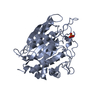
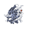
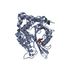
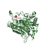
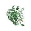
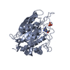
- PDB-2zef: Crystal structure of the human glutaminyl cyclase mutant E201D at... -
+ Open data

- Basic information
| Entry | Database: PDB / ID: 2zef | ||||||
|---|---|---|---|---|---|---|---|
| Title | Crystal structure of the human glutaminyl cyclase mutant E201D at 1.67 angstrom resolution | ||||||
 Components Components | Glutaminyl-peptide cyclotransferase | ||||||
 Keywords Keywords | TRANSFERASE / hydrogen bond network / glutaminyl cyclase / pyroglutamate / site-directed mutagenesis / proton transfer | ||||||
| Function / homology |  Function and homology information Function and homology informationpeptidyl-pyroglutamic acid biosynthetic process, using glutaminyl-peptide cyclotransferase / glutaminyl-peptide cyclotransferase / glutaminyl-peptide cyclotransferase activity / protein modification process / specific granule lumen / tertiary granule lumen / ficolin-1-rich granule lumen / Neutrophil degranulation / extracellular exosome / extracellular region / zinc ion binding Similarity search - Function | ||||||
| Biological species |  Homo sapiens (human) Homo sapiens (human) | ||||||
| Method |  X-RAY DIFFRACTION / SYNCHROTRON / FOURIER SYNTHESIS / Resolution: 1.67 Å X-RAY DIFFRACTION / SYNCHROTRON / FOURIER SYNTHESIS / Resolution: 1.67 Å | ||||||
 Authors Authors | Huang, K.F. / Wang, Y.R. / Chang, E.C. / Chou, T.L. / Wang, A.H. | ||||||
 Citation Citation | Journal: Biochem.J. / Year: 2008 Title: A conserved hydrogen-bond network in the catalytic centre of animal glutaminyl cyclases is critical for catalysis. Authors: Huang, K.F. / Wang, Y.R. / Chang, E.C. / Chou, T.L. / Wang, A.H. | ||||||
| History |
|
- Structure visualization
| Structure viewer | Molecule: MolmilJmol/JSmol |
|---|
- Downloads & links
-Download
| PDBx/mmCIF format | 2zef.cif.gz | 160.1 KB | Display | PDBx/mmCIF format |
|---|---|---|---|---|
| PDB format | pdb2zef.ent.gz | 124.8 KB | Display | PDB format |
| PDBx/mmJSON format | 2zef.json.gz | Tree view | PDBx/mmJSON format | |
| Others |  Other downloads Other downloads |
-Validation report
| Arichive directory | https://data.pdbj.org/pub/pdb/validation_reports/ze/2zefftp://data.pdbj.org/pub/pdb/validation_reports/ze/2zef | HTTPS FTP |
|---|
-Related structure data
| Related structure data | 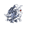 2zedC 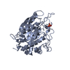 2zeeC 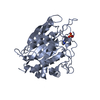 2zegC  2zehC  2zelC 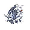 2zemC 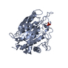 2zenC  2zeoC  2zepC  2afmS S: Starting model for refinement C: citing same article ( |
|---|---|
| Similar structure data |


-Links
 PDBj
PDBj
- Assembly
| Deposited unit | 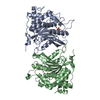
| ||||||||||||||||||
|---|---|---|---|---|---|---|---|---|---|---|---|---|---|---|---|---|---|---|---|
| 1 | 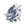
| ||||||||||||||||||
| 2 | 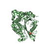
| ||||||||||||||||||
| 3 | 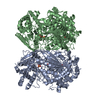
| ||||||||||||||||||
| Unit cell |
| ||||||||||||||||||
| Components on special symmetry positions |
|
-Components
| #1: Protein | Mass: 37543.371 Da / Num. of mol.: 2 / Fragment: residues 33-361 / Mutation: E201D Source method: isolated from a genetically manipulated source Source: (gene. exp.) Homo sapiens (human) / Tissue: bone marrow / Gene: QPCT / Plasmid: pET32a / Species (production host): Escherichia coli / Production host:  References: UniProt: Q16769, glutaminyl-peptide cyclotransferase #2: Chemical |   Mass: 65.409 Da / Num. of mol.: 2 / Source method: obtained synthetically / Formula: Zn Mass: 65.409 Da / Num. of mol.: 2 / Source method: obtained synthetically / Formula: Zn#3: Chemical |   Mass: 96.063 Da / Num. of mol.: 2 / Source method: obtained synthetically / Formula: SO4 Mass: 96.063 Da / Num. of mol.: 2 / Source method: obtained synthetically / Formula: SO4#4: Water | ChemComp-HOH / |  Mass: 18.015 Da / Num. of mol.: 815 / Source method: isolated from a natural source / Formula: H2O Mass: 18.015 Da / Num. of mol.: 815 / Source method: isolated from a natural source / Formula: H2O |
|---|
-Experimental details
-Experiment
| Experiment | Method: X-RAY DIFFRACTION / Number of used crystals: 1 |
|---|
- Sample preparation
| Crystal | Density Matthews: 3.03 Å3/Da / Density % sol: 59.41 % |
|---|---|
| Crystal grow | Temperature: 298 K / Method: vapor diffusion, hanging drop / pH: 6.5 Details: 2%-4% dioxane, 1.6-1.8M ammonium sulfate, 100mM Mes, pH 6.5, VAPOR DIFFUSION, HANGING DROP, temperature 298K |
-Data collection
| Diffraction | Mean temperature: 100 K |
|---|---|
| Diffraction source | Source: SYNCHROTRON / Site: SPring-8  / Beamline: BL12B2 / Wavelength: 1 Å / Beamline: BL12B2 / Wavelength: 1 Å |
| Detector | Type: ADSC QUANTUM 210 / Detector: CCD / Date: Mar 30, 2006 / Details: mirrors |
| Radiation | Monochromator: GRAPHITE / Protocol: SINGLE WAVELENGTH / Monochromatic (M) / Laue (L): M / Scattering type: x-ray |
| Radiation wavelength | Wavelength: 1 Å / Relative weight: 1 |
| Reflection | Resolution: 1.67→30 Å / Num. all: 105612 / Num. obs: 105401 / % possible obs: 99.8 % / Observed criterion σ(F): 0 / Observed criterion σ(I): 1 / Redundancy: 4.7 % / Biso Wilson estimate: 23 Å2 / Rmerge(I) obs: 0.047 / Rsym value: 0.047 / Net I/σ(I): 40 |
| Reflection shell | Resolution: 1.67→1.73 Å / Redundancy: 4.3 % / Rmerge(I) obs: 0.479 / Mean I/σ(I) obs: 3.9 / Num. unique all: 10455 / Rsym value: 0.479 / % possible all: 99.8 |
- Processing
| Software |
| |||||||||||||||||||||||||
|---|---|---|---|---|---|---|---|---|---|---|---|---|---|---|---|---|---|---|---|---|---|---|---|---|---|---|
| Refinement | Method to determine structure: FOURIER SYNTHESIS Starting model: PDB ENTRY 2AFM Resolution: 1.67→30 Å / Isotropic thermal model: Isotropic / Cross valid method: THROUGHOUT / σ(F): 0 / σ(I): 0 / Stereochemistry target values: Engh & Huber
| |||||||||||||||||||||||||
| Displacement parameters | Biso mean: 23 Å2 | |||||||||||||||||||||||||
| Refinement step | Cycle: LAST / Resolution: 1.67→30 Å
| |||||||||||||||||||||||||
| Refine LS restraints |
| |||||||||||||||||||||||||
| LS refinement shell | Resolution: 1.67→1.73 Å / Rfactor Rfree error: 0.0115
|
