 Movie
Movie Controller

[English] 日本語
 Yorodumi
- PDB-7cm0: Crystal structure of a glutaminyl cyclase in complex with NHV-1009 -
+ Open data

- Basic information
| Entry | Database: PDB / ID: 7cm0 | ||||||
|---|---|---|---|---|---|---|---|
| Title | Crystal structure of a glutaminyl cyclase in complex with NHV-1009 | ||||||
 Components Components | Glutaminyl-peptide cyclotransferase | ||||||
 Keywords Keywords | TRANSFERASE / glutaminyl cyclases (QCs) / Alzheimer disease | ||||||
| Function / homology |  Function and homology information Function and homology informationpeptidyl-pyroglutamic acid biosynthetic process, using glutaminyl-peptide cyclotransferase / glutaminyl-peptide cyclotransferase / glutaminyl-peptide cyclotransferase activity / protein modification process / specific granule lumen / tertiary granule lumen / ficolin-1-rich granule lumen / Neutrophil degranulation / extracellular exosome / extracellular region / zinc ion binding Similarity search - Function | ||||||
| Biological species |  Homo sapiens (human) Homo sapiens (human) | ||||||
| Method |  X-RAY DIFFRACTION / SYNCHROTRON / MOLECULAR REPLACEMENT / Resolution: 2.2 Å X-RAY DIFFRACTION / SYNCHROTRON / MOLECULAR REPLACEMENT / Resolution: 2.2 Å | ||||||
 Authors Authors | Lee, J.W. / Song, J.Y. / Jang, T.H. / Ha, J.H. | ||||||
 Citation Citation | Journal: Eur.J.Med.Chem. / Year: 2021 Title: Discovery of highly potent human glutaminyl cyclase (QC) inhibitors as anti-Alzheimer's agents by the combination of pharmacophore-based and structure-based design. Authors: Van Manh, N. / Hoang, V.H. / Ngo, V.T.H. / Ann, J. / Jang, T.H. / Ha, J.H. / Song, J.Y. / Ha, H.J. / Kim, H. / Kim, Y.H. / Lee, J. / Lee, J. | ||||||
| History |
|
- Structure visualization
| Structure viewer | Molecule: MolmilJmol/JSmol |
|---|
- Downloads & links
-Download
| PDBx/mmCIF format | 7cm0.cif.gz | 270 KB | Display | PDBx/mmCIF format |
|---|---|---|---|---|
| PDB format | pdb7cm0.ent.gz | 215.2 KB | Display | PDB format |
| PDBx/mmJSON format | 7cm0.json.gz | Tree view | PDBx/mmJSON format | |
| Others |  Other downloads Other downloads |
-Validation report
| Arichive directory | https://data.pdbj.org/pub/pdb/validation_reports/cm/7cm0ftp://data.pdbj.org/pub/pdb/validation_reports/cm/7cm0 | HTTPS FTP |
|---|
-Related structure data
| Related structure data |  4ywyS S: Starting model for refinement |
|---|---|
| Similar structure data |


-Links
 PDBj
PDBj
- Assembly
| Deposited unit | 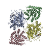
| ||||||||||||
|---|---|---|---|---|---|---|---|---|---|---|---|---|---|
| 1 | 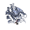
| ||||||||||||
| 2 | 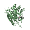
| ||||||||||||
| 3 | 
| ||||||||||||
| 4 | 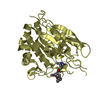
| ||||||||||||
| Unit cell |
| ||||||||||||
| Components on special symmetry positions |
|
-Components
| #1: Protein | Mass: 37399.242 Da / Num. of mol.: 4 Source method: isolated from a genetically manipulated source Source: (gene. exp.) Homo sapiens (human) / Gene: QPCT / Production host:  References: UniProt: Q16769, glutaminyl-peptide cyclotransferase #2: Chemical | ChemComp-G5R / 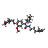  Mass: 499.646 Da / Num. of mol.: 4 / Source method: obtained synthetically / Formula: C27H41N5O4 / Feature type: SUBJECT OF INVESTIGATION Mass: 499.646 Da / Num. of mol.: 4 / Source method: obtained synthetically / Formula: C27H41N5O4 / Feature type: SUBJECT OF INVESTIGATION#3: Chemical | ChemComp-ZN /   Mass: 65.409 Da / Num. of mol.: 4 / Source method: obtained synthetically / Formula: Zn Mass: 65.409 Da / Num. of mol.: 4 / Source method: obtained synthetically / Formula: Zn#4: Water | ChemComp-HOH / |  Mass: 18.015 Da / Num. of mol.: 156 / Source method: isolated from a natural source / Formula: H2O Mass: 18.015 Da / Num. of mol.: 156 / Source method: isolated from a natural source / Formula: H2OHas ligand of interest | Y | Has protein modification | Y | |
|---|
-Experimental details
-Experiment
| Experiment | Method: X-RAY DIFFRACTION / Number of used crystals: 1 |
|---|
- Sample preparation
| Crystal | Density Matthews: 2.62 Å3/Da / Density % sol: 53.1 % |
|---|---|
| Crystal grow | Temperature: 291 K / Method: liquid diffusion / Details: 4M Ammonium acetate, pH 7.0, 0.1M Bis-Tris propane |
-Data collection
| Diffraction | Mean temperature: 100 K / Serial crystal experiment: N |
|---|---|
| Diffraction source | Source: SYNCHROTRON / Site: PAL/PLS  / Beamline: 5C (4A) / Wavelength: 0.9796 Å / Beamline: 5C (4A) / Wavelength: 0.9796 Å |
| Detector | Type: ADSC QUANTUM 315r / Detector: CCD / Date: Jul 19, 2017 |
| Radiation | Protocol: SINGLE WAVELENGTH / Monochromatic (M) / Laue (L): M / Scattering type: x-ray |
| Radiation wavelength | Wavelength: 0.9796 Å / Relative weight: 1 |
| Reflection | Resolution: 2.2→35.47 Å / Num. obs: 77174 / % possible obs: 99.46 % / Redundancy: 11.5 % / CC1/2: 0.861 / Net I/σ(I): 14.3 |
| Reflection shell | Resolution: 2.2→2.279 Å / Num. unique obs: 35430 / CC1/2: 0.861 |
- Processing
| Software |
| ||||||||||||||||||||||||
|---|---|---|---|---|---|---|---|---|---|---|---|---|---|---|---|---|---|---|---|---|---|---|---|---|---|
| Refinement | Method to determine structure: MOLECULAR REPLACEMENT Starting model: 4YWY Resolution: 2.2→33.74 Å / Cross valid method: FREE R-VALUE Stereochemistry target values: GeoStd + Monomer Library + CDL v1.2
| ||||||||||||||||||||||||
| Displacement parameters | Biso mean: 44.65 Å2 | ||||||||||||||||||||||||
| Refinement step | Cycle: LAST / Resolution: 2.2→33.74 Å
| ||||||||||||||||||||||||
| Refine LS restraints |
| ||||||||||||||||||||||||
| LS refinement shell | Resolution: 2.2→2.279 Å
|
