+ Open data
- Basic information
| Entry | Database: PDB / ID: 7cfs | |||||||||||||||||||||||||||
|---|---|---|---|---|---|---|---|---|---|---|---|---|---|---|---|---|---|---|---|---|---|---|---|---|---|---|---|---|
| Title | Cryo-EM strucutre of human acid-sensing ion channel 1a at pH 8.0 | |||||||||||||||||||||||||||
 Components Components | Acid-sensing ion channel 1 | |||||||||||||||||||||||||||
 Keywords Keywords | MEMBRANE PROTEIN / ASIC / human / resting state / cryo-EM structure | |||||||||||||||||||||||||||
| Function / homology |  Function and homology information Function and homology informationmonoatomic ion-gated channel activity / sensory perception of sour taste / pH-gated monoatomic ion channel activity / cellular response to pH / negative regulation of neurotransmitter secretion / response to acidic pH / neurotransmitter secretion / ligand-gated sodium channel activity / sodium ion transport / associative learning ...monoatomic ion-gated channel activity / sensory perception of sour taste / pH-gated monoatomic ion channel activity / cellular response to pH / negative regulation of neurotransmitter secretion / response to acidic pH / neurotransmitter secretion / ligand-gated sodium channel activity / sodium ion transport / associative learning / regulation of postsynapse assembly / behavioral fear response / sodium ion transmembrane transport / response to amphetamine / regulation of membrane potential / Stimuli-sensing channels / postsynaptic density membrane / calcium ion transmembrane transport / memory / presynapse / dendrite / glutamatergic synapse / cell surface / Golgi apparatus / plasma membrane Similarity search - Function | |||||||||||||||||||||||||||
| Biological species |  Homo sapiens (human) Homo sapiens (human) | |||||||||||||||||||||||||||
| Method | ELECTRON MICROSCOPY / single particle reconstruction / cryo EM / Resolution: 3.56 Å | |||||||||||||||||||||||||||
 Authors Authors | Sun, D.M. / Liu, S.L. / Li, S.Y. / Yang, F. / Tian, C.L. | |||||||||||||||||||||||||||
| Funding support |  China, 8items China, 8items
| |||||||||||||||||||||||||||
 Citation Citation |  Journal: Elife / Year: 2020 Journal: Elife / Year: 2020Title: Structural insights into human acid-sensing ion channel 1a inhibition by snake toxin mambalgin1. Authors: Demeng Sun / Sanling Liu / Siyu Li / Mengge Zhang / Fan Yang / Ming Wen / Pan Shi / Tao Wang / Man Pan / Shenghai Chang / Xing Zhang / Longhua Zhang / Changlin Tian / Lei Liu /  Abstract: Acid-sensing ion channels (ASICs) are proton-gated cation channels that are involved in diverse neuronal processes including pain sensing. The peptide toxin Mambalgin1 (Mamba1) from black mamba snake ...Acid-sensing ion channels (ASICs) are proton-gated cation channels that are involved in diverse neuronal processes including pain sensing. The peptide toxin Mambalgin1 (Mamba1) from black mamba snake venom can reversibly inhibit the conductance of ASICs, causing an analgesic effect. However, the detailed mechanism by which Mamba1 inhibits ASIC1s, especially how Mamba1 binding to the extracellular domain affects the conformational changes of the transmembrane domain of ASICs remains elusive. Here, we present single-particle cryo-EM structures of human ASIC1a (hASIC1a) and the hASIC1a-Mamba1 complex at resolutions of 3.56 and 3.90 Å, respectively. The structures revealed the inhibited conformation of hASIC1a upon Mamba1 binding. The combination of the structural and physiological data indicates that Mamba1 preferentially binds hASIC1a in a closed state and reduces the proton sensitivity of the channel, representing a closed-state trapping mechanism. | |||||||||||||||||||||||||||
| History |
|
- Structure visualization
| Movie |
 Movie viewer Movie viewer |
|---|---|
| Structure viewer | Molecule:  Molmil Molmil Jmol/JSmol Jmol/JSmol |
- Downloads & links
- Download
| PDBx/mmCIF format |  7cfs.cif.gz 7cfs.cif.gz | 237.6 KB | Display |  PDBx/mmCIF format PDBx/mmCIF format |
|---|---|---|---|---|
| PDB format |  pdb7cfs.ent.gz pdb7cfs.ent.gz | 187.5 KB | Display |  PDB format PDB format |
| PDBx/mmJSON format |  7cfs.json.gz 7cfs.json.gz | Tree view |  PDBx/mmJSON format PDBx/mmJSON format | |
| Others |  Other downloads Other downloads |
-Validation report
| Summary document |  7cfs_validation.pdf.gz 7cfs_validation.pdf.gz | 927.5 KB | Display |  wwPDB validaton report wwPDB validaton report |
|---|---|---|---|---|
| Full document |  7cfs_full_validation.pdf.gz 7cfs_full_validation.pdf.gz | 942.2 KB | Display | |
| Data in XML |  7cfs_validation.xml.gz 7cfs_validation.xml.gz | 40.1 KB | Display | |
| Data in CIF |  7cfs_validation.cif.gz 7cfs_validation.cif.gz | 58.9 KB | Display | |
| Arichive directory |  https://data.pdbj.org/pub/pdb/validation_reports/cf/7cfs https://data.pdbj.org/pub/pdb/validation_reports/cf/7cfs ftp://data.pdbj.org/pub/pdb/validation_reports/cf/7cfs ftp://data.pdbj.org/pub/pdb/validation_reports/cf/7cfs | HTTPS FTP |
-Related structure data
| Related structure data |  30346MC  7cftC M: map data used to model this data C: citing same article ( |
|---|---|
| Similar structure data |
- Links
Links
- Assembly
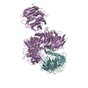
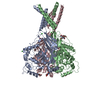
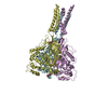
| Deposited unit | 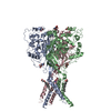
|
|---|---|
| 1 |
|
- Components
| #1: Protein | Mass: 54701.297 Da / Num. of mol.: 3 Source method: isolated from a genetically manipulated source Source: (gene. exp.)  Homo sapiens (human) / Gene: ASIC1, ACCN2, BNAC2 / Production host: Homo sapiens (human) / Gene: ASIC1, ACCN2, BNAC2 / Production host:  #2: Sugar | ChemComp-NAG / #3: Chemical | #4: Chemical | ChemComp-NA / | Has ligand of interest | N | Has protein modification | Y | |
|---|
-Experimental details
-Experiment
| Experiment | Method: ELECTRON MICROSCOPY |
|---|---|
| EM experiment | Aggregation state: PARTICLE / 3D reconstruction method: single particle reconstruction |
- Sample preparation
| Component | Name: hASIC1a / Type: COMPLEX Details: The optimized coding DNAs for human hASIC1a (Uniprot: P78348) was synthesized by GeneScript. The truncated hASIC1a (with the carboxyl terminal 60 residues removed, named as hASIC1a-DeltaC) ...Details: The optimized coding DNAs for human hASIC1a (Uniprot: P78348) was synthesized by GeneScript. The truncated hASIC1a (with the carboxyl terminal 60 residues removed, named as hASIC1a-DeltaC) was cloned into the pFastBac1 vector (Invitrogen) with 8-His tag at the amino terminus. Baculovirus-infected Sf9 cells (Thermo Fisher) were used for overexpression and were grown at 300K in serum-free SIM SF medium (Sino Biological Inc.). Entity ID: #1 / Source: RECOMBINANT |
|---|---|
| Molecular weight | Experimental value: NO |
| Source (natural) | Organism:  Homo sapiens (human) Homo sapiens (human) |
| Source (recombinant) | Organism:  Spodoptera aff. frugiperda 1 BOLD-2017 (butterflies/moths) Spodoptera aff. frugiperda 1 BOLD-2017 (butterflies/moths)Strain: Sf9 |
| Buffer solution | pH: 8 Details: 20 mM Tris (pH 8.0), 200 mM NaCl, 0.05% DDM, 0.01% CHS |
| Specimen | Conc.: 2.7 mg/ml / Embedding applied: NO / Shadowing applied: NO / Staining applied: NO / Vitrification applied: YES Details: The eluted protein in Ni-NTA affinity chromatography was further purified by size-exclusion chromatography in 20 mM Tris (pH 8.0), 200 mM NaCl, 0.05% DDM, 0.01% CHS using a Superdex200 ...Details: The eluted protein in Ni-NTA affinity chromatography was further purified by size-exclusion chromatography in 20 mM Tris (pH 8.0), 200 mM NaCl, 0.05% DDM, 0.01% CHS using a Superdex200 10/300GL column (GE HealthCare).The protein was concentrated to about 5 mg/ml based on A280 measurement, using a 100-kDa cutoff Centricon (Millipore). |
| Specimen support | Grid material: COPPER / Grid mesh size: 300 divisions/in. / Grid type: Quantifoil R1.2/1.3 |
| Vitrification | Instrument: FEI VITROBOT MARK IV / Cryogen name: ETHANE / Humidity: 100 % / Chamber temperature: 298 K Details: Purified hASIC1a-DeltaC (3 ul) at a concentration of 2.7 mg/ml was added to the freshly plasma-cleaned holey carbon grid (Quantifol, R1.2/1.3, 300 mesh, Cu), blotted for 5 s at 100% humidity ...Details: Purified hASIC1a-DeltaC (3 ul) at a concentration of 2.7 mg/ml was added to the freshly plasma-cleaned holey carbon grid (Quantifol, R1.2/1.3, 300 mesh, Cu), blotted for 5 s at 100% humidity with a Vitrobot Mark IV (ThermoFisher Scientific) and plunge frozen into liquid ethane cooled by liquid nitrogen. |
- Electron microscopy imaging
| Experimental equipment |  Model: Titan Krios / Image courtesy: FEI Company |
|---|---|
| Microscopy | Model: FEI TITAN KRIOS Details: Grids were transferred to a Titan Krios electron microscope (FEI) operated at 300 kV equipped with a Gatan K2 Summit direct detection camera. Images of hASIC1a was collected using the ...Details: Grids were transferred to a Titan Krios electron microscope (FEI) operated at 300 kV equipped with a Gatan K2 Summit direct detection camera. Images of hASIC1a was collected using the automated image acquisition software SerialEM in counting mode with 29,000x magnification yielding a pixel size of 1.014 A. The total dose of 50 e-/A2 was fractionated to 40 frames with 0.2 s per frame. Nominal defocus values ranged from -1.8 to -2.5 um. The dataset of hASIC1a included 3,235 micrographs. |
| Electron gun | Electron source:  FIELD EMISSION GUN / Accelerating voltage: 300 kV / Illumination mode: SPOT SCAN FIELD EMISSION GUN / Accelerating voltage: 300 kV / Illumination mode: SPOT SCAN |
| Electron lens | Mode: BRIGHT FIELD |
| Specimen holder | Cryogen: NITROGEN / Specimen holder model: FEI TITAN KRIOS AUTOGRID HOLDER |
| Image recording | Average exposure time: 8 sec. / Electron dose: 50 e/Å2 / Detector mode: COUNTING / Film or detector model: GATAN K2 SUMMIT (4k x 4k) / Num. of grids imaged: 2 / Num. of real images: 3235 |
| Image scans | Movie frames/image: 40 |
- Processing
| Software | Name: PHENIX / Version: 1.14_3260: / Classification: refinement | ||||||||||||||||||||||||||||
|---|---|---|---|---|---|---|---|---|---|---|---|---|---|---|---|---|---|---|---|---|---|---|---|---|---|---|---|---|---|
| EM software |
| ||||||||||||||||||||||||||||
| CTF correction | Type: PHASE FLIPPING AND AMPLITUDE CORRECTION | ||||||||||||||||||||||||||||
| Particle selection | Num. of particles selected: 274796 | ||||||||||||||||||||||||||||
| 3D reconstruction | Resolution: 3.56 Å / Resolution method: FSC 0.143 CUT-OFF / Num. of particles: 122890 / Symmetry type: POINT | ||||||||||||||||||||||||||||
| Atomic model building | Protocol: FLEXIBLE FIT | ||||||||||||||||||||||||||||
| Atomic model building | PDB-ID: 6AVE Pdb chain-ID: A / Accession code: 6AVE / Pdb chain residue range: 13-462 / Source name: PDB / Type: experimental model |
 Movie
Movie Controller




 PDBj
PDBj


