 Movie
Movie Controller

[English] 日本語
 Yorodumi
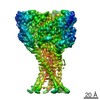
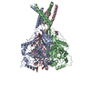
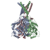
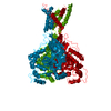
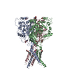
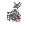
- PDB-5wky: Bromide sites in the structure of an acid sensing ion channel in ... -
+ Open data

- Basic information
| Entry | Database: PDB / ID: 5wky | ||||||||||||
|---|---|---|---|---|---|---|---|---|---|---|---|---|---|
| Title | Bromide sites in the structure of an acid sensing ion channel in a resting state | ||||||||||||
 Components Components | Acid-sensing ion channel 1 | ||||||||||||
 Keywords Keywords | TRANSPORT PROTEIN / Ion channel / ASIC / ASIC1a / Sodium channel / MEMBRANE PROTEIN | ||||||||||||
| Function / homology |  Function and homology information Function and homology informationStimuli-sensing channels / pH-gated monoatomic ion channel activity / cellular response to pH / ligand-gated sodium channel activity / sodium ion transmembrane transport / postsynaptic membrane / dendrite / glutamatergic synapse / identical protein binding / plasma membrane Similarity search - Function | ||||||||||||
| Biological species |  | ||||||||||||
| Method |  X-RAY DIFFRACTION / SYNCHROTRON / MOLECULAR REPLACEMENT / Resolution: 4 Å X-RAY DIFFRACTION / SYNCHROTRON / MOLECULAR REPLACEMENT / Resolution: 4 Å | ||||||||||||
 Authors Authors | Yoder, N. / Gouaux, E. | ||||||||||||
| Funding support |  United States, 3items United States, 3items
| ||||||||||||
 Citation Citation | Journal: PLoS ONE / Year: 2018 Title: Divalent cation and chloride ion sites of chicken acid sensing ion channel 1a elucidated by x-ray crystallography. Authors: Yoder, N. / Gouaux, E. | ||||||||||||
| History |
|
- Structure visualization
| Structure viewer | Molecule: MolmilJmol/JSmol |
|---|
- Downloads & links
-Download
| PDBx/mmCIF format | 5wky.cif.gz | 250.5 KB | Display | PDBx/mmCIF format |
|---|---|---|---|---|
| PDB format | pdb5wky.ent.gz | 195.6 KB | Display | PDB format |
| PDBx/mmJSON format | 5wky.json.gz | Tree view | PDBx/mmJSON format | |
| Others |  Other downloads Other downloads |
-Validation report
| Arichive directory | https://data.pdbj.org/pub/pdb/validation_reports/wk/5wkyftp://data.pdbj.org/pub/pdb/validation_reports/wk/5wky | HTTPS FTP |
|---|
-Related structure data
| Related structure data | 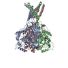 5wkxC  6cmcC  5wkuS C: citing same article ( S: Starting model for refinement |
|---|---|
| Similar structure data |


-Links
 PDBj
PDBj- Assembly
| Deposited unit | 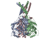
| ||||||||
|---|---|---|---|---|---|---|---|---|---|
| 1 |
| ||||||||
| Unit cell |
|
-Components
| #1: Protein | Mass: 50241.160 Da / Num. of mol.: 3 / Fragment: UNP residues 25-463 Source method: isolated from a genetically manipulated source Details: FRAGMENT: 25-463 / Source: (gene. exp.)  Homo sapiens (human) / References: UniProt: Q1XA76 Homo sapiens (human) / References: UniProt: Q1XA76#2: Polysaccharide | 2-acetamido-2-deoxy-beta-D-glucopyranose-(1-4)-2-acetamido-2-deoxy-beta-D-glucopyranose | Source method: isolated from a genetically manipulated source #3: Sugar | ChemComp-NAG /   Type: D-saccharide, beta linking / Mass: 221.208 Da / Num. of mol.: 4 Type: D-saccharide, beta linking / Mass: 221.208 Da / Num. of mol.: 4Source method: isolated from a genetically manipulated source Formula: C8H15NO6 #4: Chemical |   Mass: 35.453 Da / Num. of mol.: 3 / Source method: obtained synthetically / Formula: Cl Mass: 35.453 Da / Num. of mol.: 3 / Source method: obtained synthetically / Formula: Cl#5: Chemical | ChemComp-BA / |   Mass: 137.327 Da / Num. of mol.: 1 / Source method: obtained synthetically / Formula: Ba Mass: 137.327 Da / Num. of mol.: 1 / Source method: obtained synthetically / Formula: BaHas protein modification | Y | |
|---|
-Experimental details
-Experiment
| Experiment | Method: X-RAY DIFFRACTION / Number of used crystals: 1 |
|---|
- Sample preparation
| Crystal | Density Matthews: 3.89 Å3/Da / Density % sol: 68.37 % |
|---|---|
| Crystal grow | Temperature: 277 K / Method: vapor diffusion, hanging drop / pH: 8.5 Details: 100 mM Tris pH 8.5, 100 mM NaCl, 29% PEG 400, 5 mM BaCl2. Crystal soaked in 150 mM NaBr prior to freezing. |
-Data collection
| Diffraction | Mean temperature: 100 K |
|---|---|
| Diffraction source | Source: SYNCHROTRON / Site: APS / Beamline: 24-ID-C / Wavelength: 0.9191 Å |
| Detector | Type: DECTRIS PILATUS 6M / Detector: PIXEL / Date: Jul 14, 2016 |
| Radiation | Protocol: SINGLE WAVELENGTH / Monochromatic (M) / Laue (L): M / Scattering type: x-ray |
| Radiation wavelength | Wavelength: 0.9191 Å / Relative weight: 1 |
| Reflection | Resolution: 4→45 Å / Num. obs: 38333 / % possible obs: 99.9 % / Redundancy: 7 % / CC1/2: 1 / Net I/σ(I): 9.46 |
- Processing
| Software |
| ||||||||||||||||||||||||||||||||||||||||||||||||||||||||
|---|---|---|---|---|---|---|---|---|---|---|---|---|---|---|---|---|---|---|---|---|---|---|---|---|---|---|---|---|---|---|---|---|---|---|---|---|---|---|---|---|---|---|---|---|---|---|---|---|---|---|---|---|---|---|---|---|---|
| Refinement | Method to determine structure: MOLECULAR REPLACEMENT Starting model: 5WKU Resolution: 4→43.15 Å / SU ML: 0.53 / Cross valid method: FREE R-VALUE / σ(F): 1.34 / Phase error: 35.81 / Stereochemistry target values: ML
| ||||||||||||||||||||||||||||||||||||||||||||||||||||||||
| Solvent computation | Shrinkage radii: 0.9 Å / VDW probe radii: 1.11 Å / Solvent model: FLAT BULK SOLVENT MODEL | ||||||||||||||||||||||||||||||||||||||||||||||||||||||||
| Refinement step | Cycle: LAST / Resolution: 4→43.15 Å
| ||||||||||||||||||||||||||||||||||||||||||||||||||||||||
| Refine LS restraints |
| ||||||||||||||||||||||||||||||||||||||||||||||||||||||||
| LS refinement shell |
|
