 Movie
Movie Controller


+ Open data

- Basic information
| Entry | Database: PDB / ID: 7c7e | ||||||
|---|---|---|---|---|---|---|---|
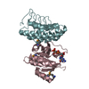
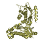
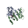
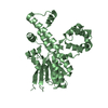
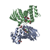
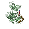
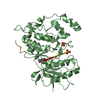
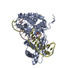
| Title | Crystal structure of C terminal domain of Escherichia coli DgoR | ||||||
 Components Components | Putative DNA-binding transcriptional regulator | ||||||
 Keywords Keywords | DNA BINDING PROTEIN / D-galactonate / GntR superfamily / transcription repressor | ||||||
| Function / homology |  Function and homology information Function and homology informationDNA-binding transcription factor activity / DNA binding / metal ion binding Similarity search - Function | ||||||
| Biological species | Escherichia coli O7:K1 | ||||||
| Method |  X-RAY DIFFRACTION / SYNCHROTRON / SAD / Resolution: 2.047 Å X-RAY DIFFRACTION / SYNCHROTRON / SAD / Resolution: 2.047 Å | ||||||
 Authors Authors | Lin, W. | ||||||
 Citation Citation | Journal: Front Microbiol / Year: 2020 Title: Structural and Functional Analyses of the Transcription Repressor DgoR From Escherichia coli Reveal a Divalent Metal-Containing D-Galactonate Binding Pocket. Authors: Lin, Z. / Sun, Y. / Liu, Y. / Tong, S. / Shang, Z. / Cai, Y. / Lin, W. | ||||||
| History |
|
- Structure visualization
| Structure viewer | Molecule: MolmilJmol/JSmol |
|---|
- Downloads & links
-Download
| PDBx/mmCIF format | 7c7e.cif.gz | 45.3 KB | Display | PDBx/mmCIF format |
|---|---|---|---|---|
| PDB format | pdb7c7e.ent.gz | 29.9 KB | Display | PDB format |
| PDBx/mmJSON format | 7c7e.json.gz | Tree view | PDBx/mmJSON format | |
| Others |  Other downloads Other downloads |
-Validation report
| Arichive directory | https://data.pdbj.org/pub/pdb/validation_reports/c7/7c7eftp://data.pdbj.org/pub/pdb/validation_reports/c7/7c7e | HTTPS FTP |
|---|
-Related structure data
| Similar structure data |
|---|


-Links
 PDBj
PDBj- Assembly
| Deposited unit | 
| ||||||||
|---|---|---|---|---|---|---|---|---|---|
| 1 | 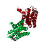
| ||||||||
| Unit cell |
|
-Components
| #1: Protein | Mass: 26485.758 Da / Num. of mol.: 1 Source method: isolated from a genetically manipulated source Source: (gene. exp.)  Gene: dgoR, ECIAI39_4297 Production host: References: UniProt: A0A0H3MRW3 |
|---|---|
| #2: Chemical | ChemComp-ZN /   Mass: 65.409 Da / Num. of mol.: 1 / Source method: obtained synthetically / Formula: Zn / Feature type: SUBJECT OF INVESTIGATION Mass: 65.409 Da / Num. of mol.: 1 / Source method: obtained synthetically / Formula: Zn / Feature type: SUBJECT OF INVESTIGATION |
| #3: Chemical | ChemComp-SO4 /   Mass: 96.063 Da / Num. of mol.: 1 / Source method: obtained synthetically / Formula: SO4 / Feature type: SUBJECT OF INVESTIGATION Mass: 96.063 Da / Num. of mol.: 1 / Source method: obtained synthetically / Formula: SO4 / Feature type: SUBJECT OF INVESTIGATION |
| #4: Chemical | ChemComp-PGE /   Mass: 150.173 Da / Num. of mol.: 1 / Source method: obtained synthetically / Formula: C6H14O4 / Feature type: SUBJECT OF INVESTIGATION Mass: 150.173 Da / Num. of mol.: 1 / Source method: obtained synthetically / Formula: C6H14O4 / Feature type: SUBJECT OF INVESTIGATION |
| #5: Water | ChemComp-HOH /  Mass: 18.015 Da / Num. of mol.: 32 / Source method: isolated from a natural source / Formula: H2O Mass: 18.015 Da / Num. of mol.: 32 / Source method: isolated from a natural source / Formula: H2O |
| Has ligand of interest | Y |
| Has protein modification | Y |
-Experimental details
-Experiment
| Experiment | Method: X-RAY DIFFRACTION / Number of used crystals: 1 |
|---|
- Sample preparation
| Crystal | Density Matthews: 2.23 Å3/Da / Density % sol: 44.9 % |
|---|---|
| Crystal grow | Temperature: 295 K / Method: vapor diffusion, hanging drop Details: 5% PEG 400, 100mM Sodium acetate pH 5.5, 2M Lithium sulfate, 100mM Magnesium sulfate |
-Data collection
| Diffraction | Mean temperature: 100 K / Serial crystal experiment: N | ||||||||||||||||||||||||||||||
|---|---|---|---|---|---|---|---|---|---|---|---|---|---|---|---|---|---|---|---|---|---|---|---|---|---|---|---|---|---|---|---|
| Diffraction source | Source: SYNCHROTRON / Site: SSRF  / Beamline: BL17U1 / Wavelength: 0.979 Å / Beamline: BL17U1 / Wavelength: 0.979 Å | ||||||||||||||||||||||||||||||
| Detector | Type: DECTRIS PILATUS3 S 6M / Detector: PIXEL / Date: Nov 27, 2019 | ||||||||||||||||||||||||||||||
| Radiation | Protocol: SINGLE WAVELENGTH / Monochromatic (M) / Laue (L): M / Scattering type: x-ray | ||||||||||||||||||||||||||||||
| Radiation wavelength | Wavelength: 0.979 Å / Relative weight: 1 | ||||||||||||||||||||||||||||||
| Reflection | Resolution: 2.04→73.65 Å / Num. obs: 15623 / % possible obs: 100 % / Redundancy: 20 % / CC1/2: 1 / Rmerge(I) obs: 0.091 / Rpim(I) all: 0.015 / Rrim(I) all: 0.093 / Net I/σ(I): 34.6 | ||||||||||||||||||||||||||||||
| Reflection shell | Diffraction-ID: 1
|
- Processing
| Software |
| ||||||||||||||||||||||||||||||||||||||||||||||||||||||||||||||||||
|---|---|---|---|---|---|---|---|---|---|---|---|---|---|---|---|---|---|---|---|---|---|---|---|---|---|---|---|---|---|---|---|---|---|---|---|---|---|---|---|---|---|---|---|---|---|---|---|---|---|---|---|---|---|---|---|---|---|---|---|---|---|---|---|---|---|---|---|
| Refinement | Method to determine structure: SAD / Resolution: 2.047→28.01 Å / SU ML: 0.26 / Cross valid method: THROUGHOUT / σ(F): 1.95 / Phase error: 28.21 / Stereochemistry target values: ML
| ||||||||||||||||||||||||||||||||||||||||||||||||||||||||||||||||||
| Solvent computation | Shrinkage radii: 0.9 Å / VDW probe radii: 1.11 Å / Solvent model: FLAT BULK SOLVENT MODEL | ||||||||||||||||||||||||||||||||||||||||||||||||||||||||||||||||||
| Displacement parameters | Biso max: 120.31 Å2 / Biso mean: 54.1432 Å2 / Biso min: 25.92 Å2 | ||||||||||||||||||||||||||||||||||||||||||||||||||||||||||||||||||
| Refinement step | Cycle: final / Resolution: 2.047→28.01 Å
| ||||||||||||||||||||||||||||||||||||||||||||||||||||||||||||||||||
| LS refinement shell | Refine-ID: X-RAY DIFFRACTION / Rfactor Rfree error: 0
|
