 Movie
Movie Controller

[English] 日本語
 Yorodumi
- PDB-1p7d: Crystal structure of the Lambda Integrase (residues 75-356) bound... -
+ Open data

- Basic information
| Entry | Database: PDB / ID: 1p7d | ||||||
|---|---|---|---|---|---|---|---|
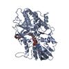
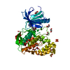
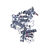
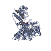
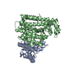
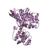
| Title | Crystal structure of the Lambda Integrase (residues 75-356) bound to DNA | ||||||
 Components Components |
| ||||||
 Keywords Keywords | DNA Binding Protein/DNA / PROTEIN-DNA COMPLEX / DNA Binding Protein-DNA COMPLEX | ||||||
| Function / homology |  Function and homology information Function and homology informationprovirus excision / integrase activity / DNA integration / viral genome integration into host DNA / establishment of integrated proviral latency / Transferases; Transferring phosphorus-containing groups; Nucleotidyltransferases / transferase activity / DNA recombination / Hydrolases; Acting on ester bonds / hydrolase activity ...provirus excision / integrase activity / DNA integration / viral genome integration into host DNA / establishment of integrated proviral latency / Transferases; Transferring phosphorus-containing groups; Nucleotidyltransferases / transferase activity / DNA recombination / Hydrolases; Acting on ester bonds / hydrolase activity / symbiont entry into host cell / DNA binding Similarity search - Function | ||||||
| Biological species |  Enterobacteria phage lambda (virus) Enterobacteria phage lambda (virus) | ||||||
| Method |  X-RAY DIFFRACTION / SYNCHROTRON / MIR / Resolution: 2.95 Å X-RAY DIFFRACTION / SYNCHROTRON / MIR / Resolution: 2.95 Å | ||||||
 Authors Authors | Aihara, H. / Kwon, H.J. / Nunes-Duby, S.E. / Landy, A. / Ellenberger, T. | ||||||
 Citation Citation | Journal: Mol.Cell / Year: 2003 Title: A Conformational Switch Controls the DNA Cleavage Activity of Lambda Integrase Authors: Aihara, H. / Kwon, H.J. / Nunes-Duby, S.E. / Landy, A. / Ellenberger, T. | ||||||
| History |
| ||||||
| Remark 999 | translational initiation |
- Structure visualization
| Structure viewer | Molecule: MolmilJmol/JSmol |
|---|
- Downloads & links
-Download
| PDBx/mmCIF format | 1p7d.cif.gz | 160.6 KB | Display | PDBx/mmCIF format |
|---|---|---|---|---|
| PDB format | pdb1p7d.ent.gz | 122.2 KB | Display | PDB format |
| PDBx/mmJSON format | 1p7d.json.gz | Tree view | PDBx/mmJSON format | |
| Others |  Other downloads Other downloads |
-Validation report
| Arichive directory | https://data.pdbj.org/pub/pdb/validation_reports/p7/1p7dftp://data.pdbj.org/pub/pdb/validation_reports/p7/1p7d | HTTPS FTP |
|---|
-Related structure data
| Similar structure data |
|---|


-Links
 PDBj
PDBj


- Assembly
| Deposited unit | 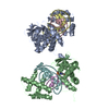
| ||||||||
|---|---|---|---|---|---|---|---|---|---|
| 1 | 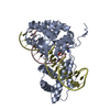
| ||||||||
| 2 | 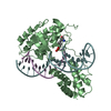
| ||||||||
| Unit cell |
|
-Components
| #1: DNA chain | Mass: 3910.573 Da / Num. of mol.: 2 / Source method: obtained synthetically #2: DNA chain | Mass: 8075.239 Da / Num. of mol.: 2 / Source method: obtained synthetically #3: Protein | Mass: 31914.553 Da / Num. of mol.: 2 / Fragment: Residues 75-356 Source method: isolated from a genetically manipulated source Source: (gene. exp.) Enterobacteria phage lambda (virus) / Genus: Lambda-like viruses / Gene: INT / Production host:  Has protein modification | Y | |
|---|
-Experimental details
-Experiment
| Experiment | Method: X-RAY DIFFRACTION / Number of used crystals: 1 |
|---|
- Sample preparation
| Crystal | Density Matthews: 2.78 Å3/Da / Density % sol: 55.42 % | ||||||||||||||||||||||||||||||||||||
|---|---|---|---|---|---|---|---|---|---|---|---|---|---|---|---|---|---|---|---|---|---|---|---|---|---|---|---|---|---|---|---|---|---|---|---|---|---|
| Crystal grow | Temperature: 295 K / Method: vapor diffusion, hanging drop / pH: 6.5 Details: PEG 8000, Ammonium Sulfate, Sodium Cacodylate, pH 6.5, VAPOR DIFFUSION, HANGING DROP, temperature 295K | ||||||||||||||||||||||||||||||||||||
| Components of the solutions |
| ||||||||||||||||||||||||||||||||||||
| Crystal grow | *PLUS Temperature: 22 ℃ / Method: vapor diffusion, hanging drop | ||||||||||||||||||||||||||||||||||||
| Components of the solutions | *PLUS
|
-Data collection
| Diffraction | Mean temperature: 100 K |
|---|---|
| Diffraction source | Source: SYNCHROTRON / Site: NSLS  / Beamline: X25 / Wavelength: 1.1 Å / Beamline: X25 / Wavelength: 1.1 Å |
| Detector | Type: BRANDEIS - B4 / Detector: CCD / Date: Oct 6, 2001 |
| Radiation | Monochromator: Si(111) / Protocol: SINGLE WAVELENGTH / Monochromatic (M) / Laue (L): M / Scattering type: x-ray |
| Radiation wavelength | Wavelength: 1.1 Å / Relative weight: 1 |
| Reflection | Resolution: 2.95→50 Å / Num. all: 19010 / Num. obs: 19010 / % possible obs: 99.8 % / Observed criterion σ(F): 0 / Observed criterion σ(I): -3 / Redundancy: 3.8 % / Biso Wilson estimate: 58.9 Å2 / Rmerge(I) obs: 0.07 / Net I/σ(I): 18.8 |
| Reflection shell | Resolution: 2.95→3.06 Å / Redundancy: 3.8 % / Rmerge(I) obs: 0.338 / Mean I/σ(I) obs: 3.6 / Num. unique all: 1884 / % possible all: 99.9 |
| Reflection | *PLUS Rmerge(I) obs: 0.07 |
| Reflection shell | *PLUS % possible obs: 99.9 % |
- Processing
| Software |
| ||||||||||||||||||||||||||||
|---|---|---|---|---|---|---|---|---|---|---|---|---|---|---|---|---|---|---|---|---|---|---|---|---|---|---|---|---|---|
| Refinement | Method to determine structure: MIR / Resolution: 2.95→50 Å / σ(F): 0 / Stereochemistry target values: Engh & Huber
| ||||||||||||||||||||||||||||
| Refinement step | Cycle: LAST / Resolution: 2.95→50 Å
| ||||||||||||||||||||||||||||
| Refine LS restraints |
| ||||||||||||||||||||||||||||
| LS refinement shell | Resolution: 2.95→3.01 Å
| ||||||||||||||||||||||||||||
| Refinement | *PLUS | ||||||||||||||||||||||||||||
| Solvent computation | *PLUS | ||||||||||||||||||||||||||||
| Displacement parameters | *PLUS | ||||||||||||||||||||||||||||
| LS refinement shell | *PLUS Rfactor Rwork: 0.401 |
