 Movie
Movie Controller

[English] 日本語
 Yorodumi
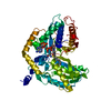
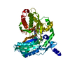
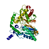
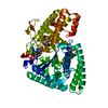
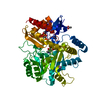
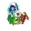
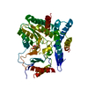
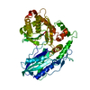
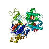
- PDB-7c1r: Crystal structure of the starter condensation domain of rhizomide... -
+ Open data

- Basic information
| Entry | Database: PDB / ID: 7c1r | ||||||
|---|---|---|---|---|---|---|---|
| Title | Crystal structure of the starter condensation domain of rhizomide synthetase RzmA mutant H140A/R148A in complex with C8-CoA | ||||||
 Components Components | Non-ribosomal peptide synthetase modules | ||||||
 Keywords Keywords | BIOSYNTHETIC PROTEIN / nonribosomal peptide synthesis / RzmA-Cs / starter condensation (Cs) domains / C8-CoA | ||||||
| Function / homology |  Function and homology information Function and homology information2,3-dihydroxybenzoate-serine ligase activity / enterobactin synthetase complex / monocarboxylic acid biosynthetic process / enterobactin biosynthetic process / Ligases; Forming carbon-nitrogen bonds; Acid-amino-acid ligases (peptide synthases) / amino acid activation for nonribosomal peptide biosynthetic process / phosphopantetheine binding / cytosol Similarity search - Function | ||||||
| Biological species |  Paraburkholderia rhizoxinica HKI 454 (bacteria) Paraburkholderia rhizoxinica HKI 454 (bacteria) | ||||||
| Method |  X-RAY DIFFRACTION / SYNCHROTRON / MOLECULAR REPLACEMENT / Resolution: 1.698 Å X-RAY DIFFRACTION / SYNCHROTRON / MOLECULAR REPLACEMENT / Resolution: 1.698 Å | ||||||
 Authors Authors | Zhong, L. / Diao, X. / Zhang, N. / Li, F.W. / Zhou, H.B. / Chen, H.N. / Ren, X. / Zhang, Y. / Wu, D. / Bian, X. | ||||||
 Citation Citation | Journal: Nat Commun / Year: 2021 Title: Engineering and elucidation of the lipoinitiation process in nonribosomal peptide biosynthesis. Authors: Zhong, L. / Diao, X. / Zhang, N. / Li, F. / Zhou, H. / Chen, H. / Bai, X. / Ren, X. / Zhang, Y. / Wu, D. / Bian, X. | ||||||
| History |
|
- Structure visualization
| Structure viewer | Molecule: MolmilJmol/JSmol |
|---|
- Downloads & links
-Download
| PDBx/mmCIF format | 7c1r.cif.gz | 205.9 KB | Display | PDBx/mmCIF format |
|---|---|---|---|---|
| PDB format | pdb7c1r.ent.gz | 159.3 KB | Display | PDB format |
| PDBx/mmJSON format | 7c1r.json.gz | Tree view | PDBx/mmJSON format | |
| Others |  Other downloads Other downloads |
-Validation report
| Arichive directory | https://data.pdbj.org/pub/pdb/validation_reports/c1/7c1rftp://data.pdbj.org/pub/pdb/validation_reports/c1/7c1r | HTTPS FTP |
|---|
-Related structure data
| Related structure data |  7c1hSC  7c1kC  7c1lC  7c1pC  7c1sC  7c1uC S: Starting model for refinement C: citing same article ( |
|---|---|
| Similar structure data |

-Links
 PDBj
PDBj


- Assembly
| Deposited unit | 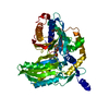
| ||||||||
|---|---|---|---|---|---|---|---|---|---|
| 1 |
| ||||||||
| Unit cell |
|
-Components
| #1: Protein | Mass: 48418.785 Da / Num. of mol.: 1 / Mutation: R148A, H140V Source method: isolated from a genetically manipulated source Details: rhizomide synthetase RzmA Source: (gene. exp.) Paraburkholderia rhizoxinica HKI 454 (bacteria)Strain: HKI 454 / Gene: RBRH_01504 / Plasmid: pET28a / Production host: References: UniProt: E5ATN9, Ligases; Forming carbon-nitrogen bonds; Acid-amino-acid ligases (peptide synthases) |
|---|---|
| #2: Chemical | ChemComp-CO8 / 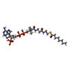  Mass: 893.730 Da / Num. of mol.: 1 / Source method: obtained synthetically / Formula: C29H50N7O17P3S / Feature type: SUBJECT OF INVESTIGATION Mass: 893.730 Da / Num. of mol.: 1 / Source method: obtained synthetically / Formula: C29H50N7O17P3S / Feature type: SUBJECT OF INVESTIGATION |
| #3: Chemical | ChemComp-TRS /   Mass: 122.143 Da / Num. of mol.: 1 / Source method: obtained synthetically / Formula: C4H12NO3 / Comment: pH buffer*YM Mass: 122.143 Da / Num. of mol.: 1 / Source method: obtained synthetically / Formula: C4H12NO3 / Comment: pH buffer*YM |
| #4: Water | ChemComp-HOH /  Mass: 18.015 Da / Num. of mol.: 377 / Source method: isolated from a natural source / Formula: H2O Mass: 18.015 Da / Num. of mol.: 377 / Source method: isolated from a natural source / Formula: H2O |
| Has ligand of interest | Y |
-Experimental details
-Experiment
| Experiment | Method: X-RAY DIFFRACTION / Number of used crystals: 1 |
|---|
- Sample preparation
| Crystal | Density Matthews: 2.13 Å3/Da / Density % sol: 42.16 % |
|---|---|
| Crystal grow | Temperature: 289 K / Method: vapor diffusion, hanging drop / pH: 9 / Details: BIS-TRIS propane, PEG 6000 |
-Data collection
| Diffraction | Mean temperature: 100 K / Serial crystal experiment: N | |||||||||||||||||||||||||||||||||||||||||||||||||||||||||||||||||||||||||||||||||||||||||||||||||||||||||||||||||||||||||||||||||||||||||||||||||||||||||||||||||||||||||||||||||||||||||||||
|---|---|---|---|---|---|---|---|---|---|---|---|---|---|---|---|---|---|---|---|---|---|---|---|---|---|---|---|---|---|---|---|---|---|---|---|---|---|---|---|---|---|---|---|---|---|---|---|---|---|---|---|---|---|---|---|---|---|---|---|---|---|---|---|---|---|---|---|---|---|---|---|---|---|---|---|---|---|---|---|---|---|---|---|---|---|---|---|---|---|---|---|---|---|---|---|---|---|---|---|---|---|---|---|---|---|---|---|---|---|---|---|---|---|---|---|---|---|---|---|---|---|---|---|---|---|---|---|---|---|---|---|---|---|---|---|---|---|---|---|---|---|---|---|---|---|---|---|---|---|---|---|---|---|---|---|---|---|---|---|---|---|---|---|---|---|---|---|---|---|---|---|---|---|---|---|---|---|---|---|---|---|---|---|---|---|---|---|---|---|---|
| Diffraction source | Source: SYNCHROTRON / Site: SSRF  / Beamline: BL19U1 / Wavelength: 0.979 Å / Beamline: BL19U1 / Wavelength: 0.979 Å | |||||||||||||||||||||||||||||||||||||||||||||||||||||||||||||||||||||||||||||||||||||||||||||||||||||||||||||||||||||||||||||||||||||||||||||||||||||||||||||||||||||||||||||||||||||||||||||
| Detector | Type: PSI PILATUS 6M / Detector: PIXEL / Date: Jan 9, 2020 / Details: mirrors | |||||||||||||||||||||||||||||||||||||||||||||||||||||||||||||||||||||||||||||||||||||||||||||||||||||||||||||||||||||||||||||||||||||||||||||||||||||||||||||||||||||||||||||||||||||||||||||
| Radiation | Monochromator: Ni FILTER / Protocol: SINGLE WAVELENGTH / Monochromatic (M) / Laue (L): M / Scattering type: x-ray | |||||||||||||||||||||||||||||||||||||||||||||||||||||||||||||||||||||||||||||||||||||||||||||||||||||||||||||||||||||||||||||||||||||||||||||||||||||||||||||||||||||||||||||||||||||||||||||
| Radiation wavelength | Wavelength: 0.979 Å / Relative weight: 1 | |||||||||||||||||||||||||||||||||||||||||||||||||||||||||||||||||||||||||||||||||||||||||||||||||||||||||||||||||||||||||||||||||||||||||||||||||||||||||||||||||||||||||||||||||||||||||||||
| Reflection | Resolution: 1.698→50 Å / Num. obs: 46330 / % possible obs: 99.8 % / Redundancy: 12.5 % / Rmerge(I) obs: 0.056 / Rpim(I) all: 0.016 / Rrim(I) all: 0.058 / Χ2: 0.962 / Net I/σ(I): 6.9 | |||||||||||||||||||||||||||||||||||||||||||||||||||||||||||||||||||||||||||||||||||||||||||||||||||||||||||||||||||||||||||||||||||||||||||||||||||||||||||||||||||||||||||||||||||||||||||||
| Reflection shell | Diffraction-ID: 1
|
- Processing
| Software |
| ||||||||||||||||||||||||||||||||||||||||||||||||||||||||||||||||||||||||||||||||||||||||||
|---|---|---|---|---|---|---|---|---|---|---|---|---|---|---|---|---|---|---|---|---|---|---|---|---|---|---|---|---|---|---|---|---|---|---|---|---|---|---|---|---|---|---|---|---|---|---|---|---|---|---|---|---|---|---|---|---|---|---|---|---|---|---|---|---|---|---|---|---|---|---|---|---|---|---|---|---|---|---|---|---|---|---|---|---|---|---|---|---|---|---|---|
| Refinement | Method to determine structure: MOLECULAR REPLACEMENT Starting model: 7C1H Resolution: 1.698→36.046 Å / SU ML: 0.17 / Cross valid method: THROUGHOUT / σ(F): 1.34 / Phase error: 22.12
| ||||||||||||||||||||||||||||||||||||||||||||||||||||||||||||||||||||||||||||||||||||||||||
| Solvent computation | Shrinkage radii: 0.9 Å / VDW probe radii: 1.11 Å | ||||||||||||||||||||||||||||||||||||||||||||||||||||||||||||||||||||||||||||||||||||||||||
| Displacement parameters | Biso max: 96.16 Å2 / Biso mean: 26.097 Å2 / Biso min: 3.83 Å2 | ||||||||||||||||||||||||||||||||||||||||||||||||||||||||||||||||||||||||||||||||||||||||||
| Refinement step | Cycle: final / Resolution: 1.698→36.046 Å
| ||||||||||||||||||||||||||||||||||||||||||||||||||||||||||||||||||||||||||||||||||||||||||
| LS refinement shell | Refine-ID: X-RAY DIFFRACTION / Rfactor Rfree error: 0
| ||||||||||||||||||||||||||||||||||||||||||||||||||||||||||||||||||||||||||||||||||||||||||
| Refinement TLS params. | Method: refined / Origin x: 11.5985 Å / Origin y: 21.9208 Å / Origin z: 24.6888 Å
| ||||||||||||||||||||||||||||||||||||||||||||||||||||||||||||||||||||||||||||||||||||||||||
| Refinement TLS group |
|
