 Movie
Movie Controller

[English] 日本語
 Yorodumi
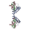
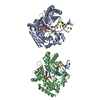
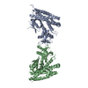
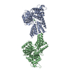
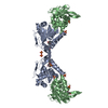
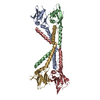
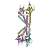
- PDB-3juv: Crystal structure of human lanosterol 14alpha-demethylase (CYP51) -
+ Open data

- Basic information
| Entry | Database: PDB / ID: 3juv | |||||||||
|---|---|---|---|---|---|---|---|---|---|---|
| Title | Crystal structure of human lanosterol 14alpha-demethylase (CYP51) | |||||||||
 Components Components | Lanosterol 14-alpha demethylase | |||||||||
 Keywords Keywords | OXIDOREDUCTASE / cytochrome p450 / sterol 14alpha-demethylase / Structural Genomics / Structural Genomics Consortium / SGC / Alternative splicing / Cholesterol biosynthesis / Endoplasmic reticulum / Heme / Iron / Lipid synthesis / Membrane / Metal-binding / Microsome / Monooxygenase / NADP / Polymorphism / Steroid biosynthesis / Sterol biosynthesis / Transmembrane | |||||||||
| Function / homology |  Function and homology information Function and homology information: / sterol 14alpha-demethylase / sterol 14-demethylase activity / negative regulation of amyloid-beta clearance / sterol metabolic process / steroid biosynthetic process / Cholesterol biosynthesis / EGR2 and SOX10-mediated initiation of Schwann cell myelination / oxidoreductase activity, acting on paired donors, with incorporation or reduction of molecular oxygen, reduced flavin or flavoprotein as one donor, and incorporation of one atom of oxygen / cholesterol biosynthetic process ...: / sterol 14alpha-demethylase / sterol 14-demethylase activity / negative regulation of amyloid-beta clearance / sterol metabolic process / steroid biosynthetic process / Cholesterol biosynthesis / EGR2 and SOX10-mediated initiation of Schwann cell myelination / oxidoreductase activity, acting on paired donors, with incorporation or reduction of molecular oxygen, reduced flavin or flavoprotein as one donor, and incorporation of one atom of oxygen / cholesterol biosynthetic process / negative regulation of protein secretion / Endogenous sterols / Activation of gene expression by SREBF (SREBP) / negative regulation of protein catabolic process / oxidoreductase activity / iron ion binding / heme binding / endoplasmic reticulum membrane / membrane Similarity search - Function | |||||||||
| Biological species |  Homo sapiens (human) Homo sapiens (human) | |||||||||
| Method |  X-RAY DIFFRACTION / SYNCHROTRON / MOLECULAR REPLACEMENT / Resolution: 3.12 Å X-RAY DIFFRACTION / SYNCHROTRON / MOLECULAR REPLACEMENT / Resolution: 3.12 Å | |||||||||
 Authors Authors | Strushkevich, N. / MacKenzie, F. / Arrowsmith, C.H. / Edwards, A.M. / Bountra, C. / Weigelt, J. / Usanov, S.A. / Park, H. / Structural Genomics Consortium (SGC) | |||||||||
 Citation Citation | Journal: J. Mol. Biol. / Year: 2010 Title: Structural basis of human CYP51 inhibition by antifungal azoles. Authors: Strushkevich, N. / Usanov, S.A. / Park, H.W. | |||||||||
| History |
|
- Structure visualization
| Structure viewer | Molecule: MolmilJmol/JSmol |
|---|
- Downloads & links
-Download
| PDBx/mmCIF format | 3juv.cif.gz | 108.4 KB | Display | PDBx/mmCIF format |
|---|---|---|---|---|
| PDB format | pdb3juv.ent.gz | 81.4 KB | Display | PDB format |
| PDBx/mmJSON format | 3juv.json.gz | Tree view | PDBx/mmJSON format | |
| Others |  Other downloads Other downloads |
-Validation report
| Arichive directory | https://data.pdbj.org/pub/pdb/validation_reports/ju/3juvftp://data.pdbj.org/pub/pdb/validation_reports/ju/3juv | HTTPS FTP |
|---|
-Related structure data
| Related structure data | 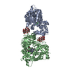 3jusC 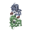 3ld6C  3i3k C: citing same article ( S: Starting model for refinement |
|---|---|
| Similar structure data |
-Links
 PDBj
PDBj


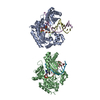
- Assembly
| Deposited unit | 
| ||||||||
|---|---|---|---|---|---|---|---|---|---|
| 1 | 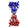
| ||||||||
| Unit cell |
|
-Components
| #1: Protein | Mass: 52769.465 Da / Num. of mol.: 1 / Fragment: UNP residues 54-502 Source method: isolated from a genetically manipulated source Source: (gene. exp.) Homo sapiens (human) / Gene: CYP51, CYP51A1 / Plasmid: pCW / Production host:  |
|---|---|
| #2: Polysaccharide | Cycloheptakis-(1-4)-(alpha-D-glucopyranose) / beta-cyclodextrin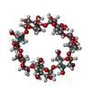  Source method: isolated from a genetically manipulated source Details: cyclic oligosaccharide / References: beta-cyclodextrin |
| #3: Chemical | ChemComp-HEM /   Mass: 616.487 Da / Num. of mol.: 1 / Source method: obtained synthetically / Formula: C34H32FeN4O4 Mass: 616.487 Da / Num. of mol.: 1 / Source method: obtained synthetically / Formula: C34H32FeN4O4 |
| #4: Water | ChemComp-HOH /  Mass: 18.015 Da / Num. of mol.: 11 / Source method: isolated from a natural source / Formula: H2O Mass: 18.015 Da / Num. of mol.: 11 / Source method: isolated from a natural source / Formula: H2O |
-Experimental details
-Experiment
| Experiment | Method: X-RAY DIFFRACTION / Number of used crystals: 1 |
|---|
- Sample preparation
| Crystal | Density Matthews: 6.75 Å3/Da / Density % sol: 81.8 % |
|---|---|
| Crystal grow | Temperature: 291 K / Method: vapor diffusion, hanging drop / pH: 7.5 Details: 2.5M (NH4)2SO4, 0.1M Hepes, pH 7.5, VAPOR DIFFUSION, HANGING DROP, temperature 291K |
-Data collection
| Diffraction | Mean temperature: 100 K |
|---|---|
| Diffraction source | Source: SYNCHROTRON / Site: APS  / Beamline: 23-ID-B / Beamline: 23-ID-B |
| Detector | Type: MARMOSAIC 300 mm CCD / Detector: CCD / Date: Jul 13, 2009 |
| Radiation | Monochromator: Si(111) / Protocol: SINGLE WAVELENGTH / Monochromatic (M) / Laue (L): M / Scattering type: x-ray |
| Radiation wavelength | Relative weight: 1 |
| Reflection | Resolution: 3.12→30 Å / Num. obs: 30272 / % possible obs: 99.1 % / Observed criterion σ(F): -3 / Observed criterion σ(I): -3 / Redundancy: 3.4 % / Biso Wilson estimate: 56.363 Å2 / Rmerge(I) obs: 0.112 / Rsym value: 0.141 / Net I/σ(I): 20.3 |
- Processing
| Software |
| ||||||||||||||||||||||||||||||||||||||||||||||||||||||||||||||||||||||||||||||||||||||||||||||||||||||||||||||||||||||||||||||||||||||||||||||||||||||||||||||||||||||||||
|---|---|---|---|---|---|---|---|---|---|---|---|---|---|---|---|---|---|---|---|---|---|---|---|---|---|---|---|---|---|---|---|---|---|---|---|---|---|---|---|---|---|---|---|---|---|---|---|---|---|---|---|---|---|---|---|---|---|---|---|---|---|---|---|---|---|---|---|---|---|---|---|---|---|---|---|---|---|---|---|---|---|---|---|---|---|---|---|---|---|---|---|---|---|---|---|---|---|---|---|---|---|---|---|---|---|---|---|---|---|---|---|---|---|---|---|---|---|---|---|---|---|---|---|---|---|---|---|---|---|---|---|---|---|---|---|---|---|---|---|---|---|---|---|---|---|---|---|---|---|---|---|---|---|---|---|---|---|---|---|---|---|---|---|---|---|---|---|---|---|---|---|
| Refinement | Method to determine structure: MOLECULAR REPLACEMENT Starting model: PDB ENTRY 3I3K 3i3k Resolution: 3.12→24.67 Å / Cor.coef. Fo:Fc: 0.945 / Cor.coef. Fo:Fc free: 0.93 / SU B: 12.825 / SU ML: 0.213 / Cross valid method: THROUGHOUT / ESU R: 0.402 / ESU R Free: 0.284 / Stereochemistry target values: MAXIMUM LIKELIHOOD / Details: HYDROGENS HAVE BEEN ADDED IN THE RIDING POSITIONS
| ||||||||||||||||||||||||||||||||||||||||||||||||||||||||||||||||||||||||||||||||||||||||||||||||||||||||||||||||||||||||||||||||||||||||||||||||||||||||||||||||||||||||||
| Solvent computation | Ion probe radii: 0.8 Å / Shrinkage radii: 0.8 Å / VDW probe radii: 1.2 Å / Solvent model: MASK | ||||||||||||||||||||||||||||||||||||||||||||||||||||||||||||||||||||||||||||||||||||||||||||||||||||||||||||||||||||||||||||||||||||||||||||||||||||||||||||||||||||||||||
| Displacement parameters | Biso mean: 75.901 Å2
| ||||||||||||||||||||||||||||||||||||||||||||||||||||||||||||||||||||||||||||||||||||||||||||||||||||||||||||||||||||||||||||||||||||||||||||||||||||||||||||||||||||||||||
| Refinement step | Cycle: LAST / Resolution: 3.12→24.67 Å
| ||||||||||||||||||||||||||||||||||||||||||||||||||||||||||||||||||||||||||||||||||||||||||||||||||||||||||||||||||||||||||||||||||||||||||||||||||||||||||||||||||||||||||
| Refine LS restraints |
| ||||||||||||||||||||||||||||||||||||||||||||||||||||||||||||||||||||||||||||||||||||||||||||||||||||||||||||||||||||||||||||||||||||||||||||||||||||||||||||||||||||||||||
| LS refinement shell | Resolution: 3.12→3.201 Å / Total num. of bins used: 20
|
