 Movie
Movie Controller

[English] 日本語
 Yorodumi
- PDB-1isr: Crystal Structure of Metabotropic Glutamate Receptor Subtype 1 Co... -
+ Open data

- Basic information
| Entry | Database: PDB / ID: 1isr | ||||||
|---|---|---|---|---|---|---|---|
| Title | Crystal Structure of Metabotropic Glutamate Receptor Subtype 1 Complexed with Glutamate and Gadolinium Ion | ||||||
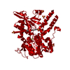
 Components Components | Metabotropic Glutamate Receptor subtype 1 | ||||||
 Keywords Keywords | SIGNALING PROTEIN / SIGNAL TRANSDUCTION / NEUROTRANSMITTER / G PROTEIN COUPLED RECEPTOR / AGONIST / GADOLINIUM ION | ||||||
| Function / homology |  Function and homology information Function and homology informationPLC activating G protein-coupled glutamate receptor activity / dendriole / : / G protein-coupled receptor dimeric complex / Class C/3 (Metabotropic glutamate/pheromone receptors) / G protein-coupled receptor homodimeric complex / : / Neurexins and neuroligins / G protein-coupled receptor activity involved in regulation of postsynaptic membrane potential / cellular response to electrical stimulus ...PLC activating G protein-coupled glutamate receptor activity / dendriole / : / G protein-coupled receptor dimeric complex / Class C/3 (Metabotropic glutamate/pheromone receptors) / G protein-coupled receptor homodimeric complex / : / Neurexins and neuroligins / G protein-coupled receptor activity involved in regulation of postsynaptic membrane potential / cellular response to electrical stimulus / synaptic signaling via neuropeptide / adenylate cyclase inhibiting G protein-coupled glutamate receptor activity / phospholipase C-activating G protein-coupled glutamate receptor signaling pathway / regulation of sensory perception of pain / neuron spine / G protein-coupled glutamate receptor signaling pathway / L-glutamate import across plasma membrane / G alpha (q) signalling events / glutamate receptor activity / regulation of glutamate secretion / synaptic transmission, GABAergic / membrane depolarization / regulation of synaptic transmission, glutamatergic / sensory perception of pain / nuclear estrogen receptor binding / calcium-mediated signaling / locomotory behavior / postsynaptic density membrane / Schaffer collateral - CA1 synapse / G protein-coupled receptor activity / presynaptic membrane / dendritic spine / phospholipase C-activating G protein-coupled receptor signaling pathway / chemical synaptic transmission / positive regulation of MAPK cascade / postsynaptic membrane / neuron projection / postsynaptic density / G protein-coupled receptor signaling pathway / axon / neuronal cell body / dendrite / glutamatergic synapse / nucleus / plasma membrane Similarity search - Function | ||||||
| Biological species |  | ||||||
| Method |  X-RAY DIFFRACTION / SYNCHROTRON / MOLECULAR REPLACEMENT / Resolution: 4 Å X-RAY DIFFRACTION / SYNCHROTRON / MOLECULAR REPLACEMENT / Resolution: 4 Å | ||||||
 Authors Authors | Tsuchiya, D. / Kunishima, N. / Kamiya, N. / Jingami, H. / Morikawa, K. | ||||||
 Citation Citation | Journal: Proc.Natl.Acad.Sci.USA / Year: 2002 Title: Structural views of the ligand-binding cores of a metabotropic glutamate receptor complexed with an antagonist and both glutamate and Gd3+. Authors: Tsuchiya, D. / Kunishima, N. / Kamiya, N. / Jingami, H. / Morikawa, K. #1: Journal: Nature / Year: 2000Title: Structural Basis of Glutamate Recognition by a Dimeric Metabotropic Glutamate Receptor Authors: Kunishima, N. / Shimada, Y. / Tsuji, Y. / Sato, T. / Yamamoto, M. / Kumasaka, T. / Nakanishi, S. / Jingami, H. / Morikawa, K. | ||||||
| History |
|
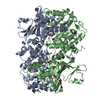
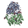
- Structure visualization
| Structure viewer | Molecule: MolmilJmol/JSmol |
|---|
- Downloads & links
-Download
| PDBx/mmCIF format | 1isr.cif.gz | 92.5 KB | Display | PDBx/mmCIF format |
|---|---|---|---|---|
| PDB format | pdb1isr.ent.gz | 67.6 KB | Display | PDB format |
| PDBx/mmJSON format | 1isr.json.gz | Tree view | PDBx/mmJSON format | |
| Others |  Other downloads Other downloads |
-Validation report
| Arichive directory | https://data.pdbj.org/pub/pdb/validation_reports/is/1isrftp://data.pdbj.org/pub/pdb/validation_reports/is/1isr | HTTPS FTP |
|---|
-Related structure data
| Related structure data |  1issC  1ewkS S: Starting model for refinement C: citing same article ( |
|---|---|
| Similar structure data |


-Links
 PDBj
PDBj


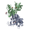
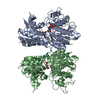
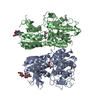
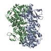
- Assembly
| Deposited unit | 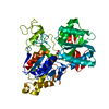
| ||||||||
|---|---|---|---|---|---|---|---|---|---|
| 1 | 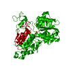
| ||||||||
| Unit cell |
| ||||||||
| Components on special symmetry positions |
| ||||||||
| Details | The biological dimer is generated by the two fold axis: x-y,-y,4/3-z |
-Components
| #1: Protein | Mass: 55258.707 Da / Num. of mol.: 1 / Fragment: EXTRACELLULAR LIGAND BINDING REGION Source method: isolated from a genetically manipulated source Source: (gene. exp.)   Spodoptera frugiperda (fall armyworm) / Strain (production host): SF9 / References: UniProt: P23385 Spodoptera frugiperda (fall armyworm) / Strain (production host): SF9 / References: UniProt: P23385 | ||
|---|---|---|---|
| #2: Chemical | ChemComp-GLU /   Type: L-peptide linking / Mass: 147.129 Da / Num. of mol.: 1 / Source method: obtained synthetically / Formula: C5H9NO4 Type: L-peptide linking / Mass: 147.129 Da / Num. of mol.: 1 / Source method: obtained synthetically / Formula: C5H9NO4 | ||
| #3: Chemical |   Mass: 157.250 Da / Num. of mol.: 2 / Source method: obtained synthetically / Formula: Gd Mass: 157.250 Da / Num. of mol.: 2 / Source method: obtained synthetically / Formula: GdHas protein modification | Y | |
-Experimental details
-Experiment
| Experiment | Method: X-RAY DIFFRACTION / Number of used crystals: 4 |
|---|
- Sample preparation
| Crystal | Density Matthews: 4.23 Å3/Da / Density % sol: 70.95 % | |||||||||||||||||||||||||||||||||||
|---|---|---|---|---|---|---|---|---|---|---|---|---|---|---|---|---|---|---|---|---|---|---|---|---|---|---|---|---|---|---|---|---|---|---|---|---|
| Crystal grow | Temperature: 293 K / Method: vapor diffusion, hanging drop / pH: 7.5 Details: PEG4000, HEPES, L-glutamate, gadolinium chloride, pH 7.5, VAPOR DIFFUSION, HANGING DROP, temperature 293K | |||||||||||||||||||||||||||||||||||
| Crystal grow | *PLUS Details: Kunishima, N., (2000) Nature, 407, 971. | |||||||||||||||||||||||||||||||||||
| Components of the solutions | *PLUS
|
-Data collection
| Diffraction | Mean temperature: 298 K |
|---|---|
| Diffraction source | Source: SYNCHROTRON / Site: SPring-8  / Beamline: BL40B2 / Wavelength: 1 Å / Beamline: BL40B2 / Wavelength: 1 Å |
| Detector | Type: ADSC QUANTUM 4 / Detector: CCD / Date: Mar 14, 2001 |
| Radiation | Monochromator: GRAPHITE / Protocol: SINGLE WAVELENGTH / Monochromatic (M) / Laue (L): M / Scattering type: x-ray |
| Radiation wavelength | Wavelength: 1 Å / Relative weight: 1 |
| Reflection | Resolution: 4→30 Å / Num. all: 5935 / Num. obs: 5935 / % possible obs: 90.1 % / Observed criterion σ(F): 0 / Observed criterion σ(I): 0 / Redundancy: 5 % / Biso Wilson estimate: 41.4 Å2 / Rmerge(I) obs: 0.101 |
| Reflection shell | Resolution: 4→4.2 Å / Redundancy: 2.8 % / Rmerge(I) obs: 0.23 / Mean I/σ(I) obs: 2.9 / Num. unique all: 440 / % possible all: 90.1 |
| Reflection | *PLUS Rmerge(I) obs: 0.1 |
| Reflection shell | *PLUS Highest resolution: 4 Å / Lowest resolution: 4.2 Å / % possible obs: 75.6 % / Rmerge(I) obs: 0.23 |
- Processing
| Software |
| |||||||||||||||||||||||||
|---|---|---|---|---|---|---|---|---|---|---|---|---|---|---|---|---|---|---|---|---|---|---|---|---|---|---|
| Refinement | Method to determine structure: MOLECULAR REPLACEMENT Starting model: CHAIN A of PDB ENTRY 1EWK Resolution: 4→20 Å / Isotropic thermal model: Isotropic / Cross valid method: THROUGHOUT / σ(F): 0 / Stereochemistry target values: Engh & Huber
| |||||||||||||||||||||||||
| Displacement parameters | Biso mean: 62.3 Å2
| |||||||||||||||||||||||||
| Refine analyze |
| |||||||||||||||||||||||||
| Refinement step | Cycle: LAST / Resolution: 4→20 Å
| |||||||||||||||||||||||||
| Refine LS restraints |
| |||||||||||||||||||||||||
| LS refinement shell | Resolution: 4→4.13 Å
| |||||||||||||||||||||||||
| Refinement | *PLUS Highest resolution: 4 Å / Lowest resolution: 12 Å / % reflection Rfree: 7 % / Rfactor obs: 0.237 | |||||||||||||||||||||||||
| Solvent computation | *PLUS | |||||||||||||||||||||||||
| Displacement parameters | *PLUS | |||||||||||||||||||||||||
| Refine LS restraints | *PLUS Type: c_bond_d / Dev ideal: 0.003 | |||||||||||||||||||||||||
| LS refinement shell | *PLUS Rfactor obs: 0.277 |
