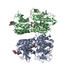
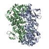
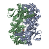
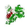
Entry Database : PDB / ID : 3lmkTitle Ligand Binding Domain of Metabotropoc glutamate receptor mGluR5 complexed with glutamate Metabotropic glutamate receptor 5 Keywords / / / / / / / / / / / / / / Function / homology Function Domain/homology Component
/ / / / / / / / / / / / / / / / / / / / / / / / / / / / / / / / / / / / / / / / / / / / / / / / / / / / / / / / / / / / / / / / / / / / / / / / / / / / / / / / / / / / / / / / / / / / / / / Biological species Homo sapiens (human)Method / / / Resolution : 2.44 Å Authors Dobrovetsky, E. / Khutoreskaya, G. / Seitova, A. / Cossar, D. / Edwards, A.M. / Arrowsmith, C.H. / Bountra, C. / Weigelt, J. / Bochkarev, A. / Structural Genomics Consortium (SGC) Journal : To be Published Title : Metabotropic Glutamate receptor mGluR5 complexed with glutamateAuthors : Dobrovetsky, E. / Khutoreskaya, G. / Seitova, A. / Cossar, D. / Edwards, A.M. / Arrowsmith, C.H. / Bountra, C. / Weigelt, J. / Bochkarev, A. History Deposition Jan 30, 2010 Deposition site / Processing site Revision 1.0 Feb 16, 2010 Provider / Type Revision 1.1 Jul 13, 2011 Group / Refinement description / Version format complianceRevision 1.2 Nov 1, 2017 Group / Category Revision 1.3 Jul 29, 2020 Group Advisory / Data collection ... Advisory / Data collection / Database references / Derived calculations / Structure summary Category chem_comp / database_PDB_caveat ... chem_comp / database_PDB_caveat / entity / pdbx_chem_comp_identifier / pdbx_entity_nonpoly / pdbx_struct_conn_angle / struct_conn / struct_ref_seq_dif / struct_site / struct_site_gen Item _chem_comp.name / _chem_comp.type ... _chem_comp.name / _chem_comp.type / _entity.pdbx_description / _pdbx_entity_nonpoly.name / _pdbx_struct_conn_angle.ptnr1_auth_comp_id / _pdbx_struct_conn_angle.ptnr1_auth_seq_id / _pdbx_struct_conn_angle.ptnr1_label_asym_id / _pdbx_struct_conn_angle.ptnr1_label_comp_id / _pdbx_struct_conn_angle.ptnr1_label_seq_id / _pdbx_struct_conn_angle.ptnr3_auth_comp_id / _pdbx_struct_conn_angle.ptnr3_auth_seq_id / _pdbx_struct_conn_angle.ptnr3_label_asym_id / _pdbx_struct_conn_angle.ptnr3_label_comp_id / _pdbx_struct_conn_angle.ptnr3_label_seq_id / _pdbx_struct_conn_angle.value / _struct_conn.pdbx_dist_value / _struct_conn.pdbx_leaving_atom_flag / _struct_conn.pdbx_role / _struct_conn.ptnr1_auth_asym_id / _struct_conn.ptnr1_auth_comp_id / _struct_conn.ptnr1_auth_seq_id / _struct_conn.ptnr1_label_asym_id / _struct_conn.ptnr1_label_atom_id / _struct_conn.ptnr1_label_comp_id / _struct_conn.ptnr1_label_seq_id / _struct_conn.ptnr2_auth_asym_id / _struct_conn.ptnr2_auth_comp_id / _struct_conn.ptnr2_auth_seq_id / _struct_conn.ptnr2_label_asym_id / _struct_conn.ptnr2_label_atom_id / _struct_conn.ptnr2_label_comp_id / _struct_conn.ptnr2_label_seq_id / _struct_ref_seq_dif.details Description / Provider / Type Revision 1.4 Oct 13, 2021 Group / Structure summary / Category / database_2 / struct_ref_seq_difItem _chem_comp.pdbx_synonyms / _database_2.pdbx_DOI ... _chem_comp.pdbx_synonyms / _database_2.pdbx_DOI / _database_2.pdbx_database_accession / _struct_ref_seq_dif.details Revision 1.5 Nov 6, 2024 Group / Structure summaryCategory chem_comp_atom / chem_comp_bond ... chem_comp_atom / chem_comp_bond / pdbx_entry_details / pdbx_modification_feature
Show all Show less
 Movie
Movie Controller

 Yorodumi
 Open data
 Basic information
 Components
 Keywords
 Function and homology information
 Homo sapiens (human)
 X-RAY DIFFRACTION /
  Authors
 Citation
 Structure visualization
 Downloads & links
 Other downloads
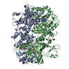


 PDBj
PDBj


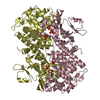
 Assembly
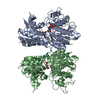
 unidentified baculovirus / References: UniProt: P41594
 Type: D-saccharide, beta linking / Mass: 221.208 Da / Num. of mol.: 2
 Type: L-peptide linking / Mass: 147.129 Da / Num. of mol.: 2 / Source method: obtained synthetically / Formula: C5H9NO4
 Mass: 24.305 Da / Num. of mol.: 3 / Source method: obtained synthetically / Formula: Mg
 Mass: 18.015 Da / Num. of mol.: 89 / Source method: isolated from a natural source / Formula: H2O
 Sample preparation
 Processing