 Movie
Movie Controller

[English] 日本語
 Yorodumi
- PDB-7bv3: Crystal structure of a ugt transferase from Siraitia grosvenorii ... -
+ Open data

- Basic information
| Entry | Database: PDB / ID: 7bv3 | ||||||
|---|---|---|---|---|---|---|---|
| Title | Crystal structure of a ugt transferase from Siraitia grosvenorii in complex with UDP | ||||||
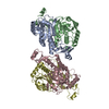
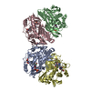
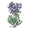
 Components Components | Glycosyltransferase | ||||||
 Keywords Keywords | TRANSFERASE / prenyltransferase | ||||||
| Function / homology |  Function and homology information Function and homology informationUDP-glycosyltransferase activity / Transferases; Glycosyltransferases; Hexosyltransferases / chloroplast / nucleotide binding Similarity search - Function | ||||||
| Biological species |  Siraitia grosvenorii (monk fruit) Siraitia grosvenorii (monk fruit) | ||||||
| Method |  X-RAY DIFFRACTION / SYNCHROTRON / MOLECULAR REPLACEMENT / Resolution: 1.85 Å X-RAY DIFFRACTION / SYNCHROTRON / MOLECULAR REPLACEMENT / Resolution: 1.85 Å | ||||||
 Authors Authors | Li, J. / Shan, N. / Yang, J.G. / Liu, W.D. / Sun, Y.X. | ||||||
 Citation Citation | Journal: Green Synth Catal / Year: 2021 Title: Near-perfect control of the regioselective glucosylation enabled by rational design of glycosyltransferases Authors: Li, J. / Qu, G. / Shang, N. / Chen, P. / Men, Y. / Liu, W.D. / Mei, Z. / Sun, Y.X. / Sun, Z. | ||||||
| History |
|
- Structure visualization
| Structure viewer | Molecule: MolmilJmol/JSmol |
|---|
- Downloads & links
-Download
| PDBx/mmCIF format | 7bv3.cif.gz | 200.7 KB | Display | PDBx/mmCIF format |
|---|---|---|---|---|
| PDB format | pdb7bv3.ent.gz | 156.8 KB | Display | PDB format |
| PDBx/mmJSON format | 7bv3.json.gz | Tree view | PDBx/mmJSON format | |
| Others |  Other downloads Other downloads |
-Validation report
| Arichive directory | https://data.pdbj.org/pub/pdb/validation_reports/bv/7bv3ftp://data.pdbj.org/pub/pdb/validation_reports/bv/7bv3 | HTTPS FTP |
|---|
-Related structure data
| Related structure data |  2acwS S: Starting model for refinement |
|---|---|
| Similar structure data |


-Links
 PDBj
PDBj


- Assembly
| Deposited unit | 
| ||||||||
|---|---|---|---|---|---|---|---|---|---|
| 1 |
| ||||||||
| Unit cell |
| ||||||||
| Components on special symmetry positions |
|
-Components
| #1: Protein | Mass: 52805.641 Da / Num. of mol.: 2 Source method: isolated from a genetically manipulated source Source: (gene. exp.) Siraitia grosvenorii (monk fruit) / Gene: UGT74AC2 / Plasmid: pET-28a / Production host:  References: UniProt: A0A346A6C4, Transferases; Glycosyltransferases; Hexosyltransferases #2: Chemical |   Type: RNA linking / Mass: 404.161 Da / Num. of mol.: 2 / Source method: obtained synthetically / Formula: C9H14N2O12P2 / Feature type: SUBJECT OF INVESTIGATION / Comment: UDP*YM Type: RNA linking / Mass: 404.161 Da / Num. of mol.: 2 / Source method: obtained synthetically / Formula: C9H14N2O12P2 / Feature type: SUBJECT OF INVESTIGATION / Comment: UDP*YM#3: Water | ChemComp-HOH / |  Mass: 18.015 Da / Num. of mol.: 656 / Source method: isolated from a natural source / Formula: H2O Mass: 18.015 Da / Num. of mol.: 656 / Source method: isolated from a natural source / Formula: H2OHas ligand of interest | Y | |
|---|
-Experimental details
-Experiment
| Experiment | Method: X-RAY DIFFRACTION / Number of used crystals: 1 |
|---|
- Sample preparation
| Crystal | Density Matthews: 2.74 Å3/Da / Density % sol: 55.05 % / Mosaicity: 0.243 ° |
|---|---|
| Crystal grow | Temperature: 295 K / Method: vapor diffusion, sitting drop / pH: 8 / Details: PEG8000, MgAc |
-Data collection
| Diffraction | Mean temperature: 100 K / Serial crystal experiment: N | |||||||||||||||||||||||||||||||||||||||||||||||||||||||||||||||||||||||||||||||||||||||||||||||||||
|---|---|---|---|---|---|---|---|---|---|---|---|---|---|---|---|---|---|---|---|---|---|---|---|---|---|---|---|---|---|---|---|---|---|---|---|---|---|---|---|---|---|---|---|---|---|---|---|---|---|---|---|---|---|---|---|---|---|---|---|---|---|---|---|---|---|---|---|---|---|---|---|---|---|---|---|---|---|---|---|---|---|---|---|---|---|---|---|---|---|---|---|---|---|---|---|---|---|---|---|---|
| Diffraction source | Source: SYNCHROTRON / Site: SSRF  / Beamline: BL19U1 / Wavelength: 1 Å / Beamline: BL19U1 / Wavelength: 1 Å | |||||||||||||||||||||||||||||||||||||||||||||||||||||||||||||||||||||||||||||||||||||||||||||||||||
| Detector | Type: DECTRIS PILATUS 2M / Detector: PIXEL / Date: Dec 5, 2019 | |||||||||||||||||||||||||||||||||||||||||||||||||||||||||||||||||||||||||||||||||||||||||||||||||||
| Radiation | Monochromator: GRAPHITE / Protocol: SINGLE WAVELENGTH / Monochromatic (M) / Laue (L): M / Scattering type: x-ray | |||||||||||||||||||||||||||||||||||||||||||||||||||||||||||||||||||||||||||||||||||||||||||||||||||
| Radiation wavelength | Wavelength: 1 Å / Relative weight: 1 | |||||||||||||||||||||||||||||||||||||||||||||||||||||||||||||||||||||||||||||||||||||||||||||||||||
| Reflection | Resolution: 1.85→25 Å / Num. obs: 98386 / % possible obs: 99.9 % / Redundancy: 6.6 % / Rmerge(I) obs: 0.062 / Rpim(I) all: 0.026 / Rrim(I) all: 0.068 / Χ2: 0.711 / Net I/σ(I): 8.8 / Num. measured all: 651819 | |||||||||||||||||||||||||||||||||||||||||||||||||||||||||||||||||||||||||||||||||||||||||||||||||||
| Reflection shell | Diffraction-ID: 1
|
- Processing
| Software |
| |||||||||||||||||||||||||||||||||||||||||||||||||||||||
|---|---|---|---|---|---|---|---|---|---|---|---|---|---|---|---|---|---|---|---|---|---|---|---|---|---|---|---|---|---|---|---|---|---|---|---|---|---|---|---|---|---|---|---|---|---|---|---|---|---|---|---|---|---|---|---|---|
| Refinement | Method to determine structure: MOLECULAR REPLACEMENT Starting model: 2ACW Resolution: 1.85→24.22 Å / SU ML: 0.16 / Cross valid method: THROUGHOUT / σ(F): 1.34 / Phase error: 20.21 / Stereochemistry target values: ML
| |||||||||||||||||||||||||||||||||||||||||||||||||||||||
| Solvent computation | Shrinkage radii: 0.9 Å / VDW probe radii: 1.11 Å / Solvent model: FLAT BULK SOLVENT MODEL | |||||||||||||||||||||||||||||||||||||||||||||||||||||||
| Displacement parameters | Biso max: 128.37 Å2 / Biso mean: 38.3874 Å2 / Biso min: 11.94 Å2 | |||||||||||||||||||||||||||||||||||||||||||||||||||||||
| Refinement step | Cycle: final / Resolution: 1.85→24.22 Å
| |||||||||||||||||||||||||||||||||||||||||||||||||||||||
| LS refinement shell | Refine-ID: X-RAY DIFFRACTION / Rfactor Rfree error: 0 / Num. reflection Rfree: 490 / Total num. of bins used: 10 / % reflection obs: 100 %
|
