 Movie
Movie Controller


+ Open data

- Basic information
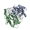
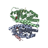
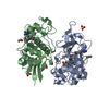
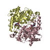
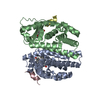
| Entry | Database: PDB / ID: 3euf | ||||||
|---|---|---|---|---|---|---|---|
| Title | Crystal structure of BAU-bound human uridine phosphorylase 1 | ||||||
 Components Components | Uridine phosphorylase 1 | ||||||
 Keywords Keywords | TRANSFERASE / nucleoside phosphorylase / uridine rescue / 5-benzylacyclouridine / Alternative splicing / Glycosyltransferase | ||||||
| Function / homology |  Function and homology information Function and homology informationCMP catabolic process / deoxyuridine phosphorylase activity / dTMP catabolic process / dCMP catabolic process / thymidine phosphorylase activity / UMP catabolic process / dUMP catabolic process / uridine catabolic process / Pyrimidine salvage / Pyrimidine catabolism ...CMP catabolic process / deoxyuridine phosphorylase activity / dTMP catabolic process / dCMP catabolic process / thymidine phosphorylase activity / UMP catabolic process / dUMP catabolic process / uridine catabolic process / Pyrimidine salvage / Pyrimidine catabolism / uridine phosphorylase / UMP salvage / uridine phosphorylase activity / nucleobase-containing compound metabolic process / cellular response to glucose starvation / nucleoplasm / identical protein binding / cytosol Similarity search - Function | ||||||
| Biological species |  Homo sapiens (human) Homo sapiens (human) | ||||||
| Method |  X-RAY DIFFRACTION / SYNCHROTRON / MOLECULAR REPLACEMENT / Resolution: 1.9 Å X-RAY DIFFRACTION / SYNCHROTRON / MOLECULAR REPLACEMENT / Resolution: 1.9 Å | ||||||
 Authors Authors | Roosild, T.P. | ||||||
 Citation Citation | Journal: Bmc Struct.Biol. / Year: 2009 Title: Implications of the structure of human uridine phosphorylase 1 on the development of novel inhibitors for improving the therapeutic window of fluoropyrimidine chemotherapy. Authors: Roosild, T.P. / Castronovo, S. / Fabbiani, M. / Pizzorno, G. | ||||||
| History |
|
- Structure visualization
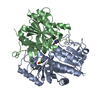
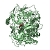
| Structure viewer | Molecule: MolmilJmol/JSmol |
|---|
- Downloads & links
-Download
| PDBx/mmCIF format | 3euf.cif.gz | 240.2 KB | Display | PDBx/mmCIF format |
|---|---|---|---|---|
| PDB format | pdb3euf.ent.gz | 195.1 KB | Display | PDB format |
| PDBx/mmJSON format | 3euf.json.gz | Tree view | PDBx/mmJSON format | |
| Others |  Other downloads Other downloads |
-Validation report
| Arichive directory | https://data.pdbj.org/pub/pdb/validation_reports/eu/3eufftp://data.pdbj.org/pub/pdb/validation_reports/eu/3euf | HTTPS FTP |
|---|
-Related structure data
| Related structure data |  3eueC  1u1cS C: citing same article ( S: Starting model for refinement |
|---|---|
| Similar structure data |


-Links
 PDBj
PDBj- Assembly
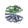
| Deposited unit | 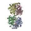
| ||||||||||||||||||||||||||||||||||||||||||||||||||||||||||||||||||
|---|---|---|---|---|---|---|---|---|---|---|---|---|---|---|---|---|---|---|---|---|---|---|---|---|---|---|---|---|---|---|---|---|---|---|---|---|---|---|---|---|---|---|---|---|---|---|---|---|---|---|---|---|---|---|---|---|---|---|---|---|---|---|---|---|---|---|---|
| 1 | 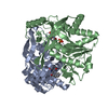
| ||||||||||||||||||||||||||||||||||||||||||||||||||||||||||||||||||
| 2 | 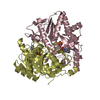
| ||||||||||||||||||||||||||||||||||||||||||||||||||||||||||||||||||
| Unit cell |
| ||||||||||||||||||||||||||||||||||||||||||||||||||||||||||||||||||
| Noncrystallographic symmetry (NCS) | NCS domain:
NCS domain segments:
NCS ensembles :
|
-Components
| #1: Protein | Mass: 36050.617 Da / Num. of mol.: 4 Source method: isolated from a genetically manipulated source Source: (gene. exp.) Homo sapiens (human) / Gene: UPP1, UP / Plasmid: pQE / Production host:  #2: Chemical | ChemComp-BAU / 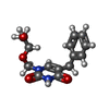  Mass: 276.288 Da / Num. of mol.: 4 / Source method: obtained synthetically / Formula: C14H16N2O4 Mass: 276.288 Da / Num. of mol.: 4 / Source method: obtained synthetically / Formula: C14H16N2O4#3: Chemical | ChemComp-PO4 /   Mass: 94.971 Da / Num. of mol.: 4 / Source method: obtained synthetically / Formula: PO4 Mass: 94.971 Da / Num. of mol.: 4 / Source method: obtained synthetically / Formula: PO4#4: Water | ChemComp-HOH / |  Mass: 18.015 Da / Num. of mol.: 336 / Source method: isolated from a natural source / Formula: H2O Mass: 18.015 Da / Num. of mol.: 336 / Source method: isolated from a natural source / Formula: H2O |
|---|
-Experimental details
-Experiment
| Experiment | Method: X-RAY DIFFRACTION / Number of used crystals: 1 |
|---|
- Sample preparation
| Crystal | Density Matthews: 2.24 Å3/Da / Density % sol: 45.2 % |
|---|---|
| Crystal grow | Temperature: 298 K / Method: vapor diffusion, hanging drop / pH: 5.5 Details: 17% PEG 3350, 300mM KCl, 30mM MgCl2, 100mM Bis-Tris Buffer, pH 5.5, VAPOR DIFFUSION, HANGING DROP, temperature 298K |
-Data collection
| Diffraction | Mean temperature: 100 K |
|---|---|
| Diffraction source | Source: SYNCHROTRON / Site: SSRL  / Beamline: BL7-1 / Wavelength: 1 Å / Beamline: BL7-1 / Wavelength: 1 Å |
| Detector | Type: ADSC QUANTUM 315 / Detector: CCD / Date: Apr 10, 2008 |
| Radiation | Protocol: SINGLE WAVELENGTH / Monochromatic (M) / Laue (L): M / Scattering type: x-ray |
| Radiation wavelength | Wavelength: 1 Å / Relative weight: 1 |
| Reflection | Resolution: 1.9→50 Å / Num. all: 103357 / Num. obs: 103213 / % possible obs: 99.8 % / Redundancy: 10.6 % / Rmerge(I) obs: 0.119 / Net I/σ(I): 16.1 |
| Reflection shell | Resolution: 1.9→1.97 Å / % possible obs: 100 % / Rmerge(I) obs: 0.441 / Mean I/σ(I) obs: 2 / Num. unique all: 10192 / Num. unique obs: 10192 / % possible all: 100 |
- Processing
| Software |
| ||||||||||||||||||||||||||||||||||||||||||||||||||||||||||||||||||||||||||||||||||||||||||||||||||||||||||||||||||||||||||||||||||||||||||||||||||||||||||||||||||||||||||
|---|---|---|---|---|---|---|---|---|---|---|---|---|---|---|---|---|---|---|---|---|---|---|---|---|---|---|---|---|---|---|---|---|---|---|---|---|---|---|---|---|---|---|---|---|---|---|---|---|---|---|---|---|---|---|---|---|---|---|---|---|---|---|---|---|---|---|---|---|---|---|---|---|---|---|---|---|---|---|---|---|---|---|---|---|---|---|---|---|---|---|---|---|---|---|---|---|---|---|---|---|---|---|---|---|---|---|---|---|---|---|---|---|---|---|---|---|---|---|---|---|---|---|---|---|---|---|---|---|---|---|---|---|---|---|---|---|---|---|---|---|---|---|---|---|---|---|---|---|---|---|---|---|---|---|---|---|---|---|---|---|---|---|---|---|---|---|---|---|---|---|---|
| Refinement | Method to determine structure: MOLECULAR REPLACEMENT Starting model: PDB ENTRY 1U1C Resolution: 1.9→50 Å / Cor.coef. Fo:Fc: 0.952 / Cor.coef. Fo:Fc free: 0.929 / SU B: 3.403 / SU ML: 0.102 / Cross valid method: THROUGHOUT / ESU R: 0.155 / ESU R Free: 0.151 / Stereochemistry target values: MAXIMUM LIKELIHOOD / Details: HYDROGENS HAVE BEEN ADDED IN THE RIDING POSITIONS
| ||||||||||||||||||||||||||||||||||||||||||||||||||||||||||||||||||||||||||||||||||||||||||||||||||||||||||||||||||||||||||||||||||||||||||||||||||||||||||||||||||||||||||
| Solvent computation | Ion probe radii: 0.8 Å / Shrinkage radii: 0.8 Å / VDW probe radii: 1.2 Å / Solvent model: MASK | ||||||||||||||||||||||||||||||||||||||||||||||||||||||||||||||||||||||||||||||||||||||||||||||||||||||||||||||||||||||||||||||||||||||||||||||||||||||||||||||||||||||||||
| Displacement parameters | Biso mean: 29.999 Å2
| ||||||||||||||||||||||||||||||||||||||||||||||||||||||||||||||||||||||||||||||||||||||||||||||||||||||||||||||||||||||||||||||||||||||||||||||||||||||||||||||||||||||||||
| Refinement step | Cycle: LAST / Resolution: 1.9→50 Å
| ||||||||||||||||||||||||||||||||||||||||||||||||||||||||||||||||||||||||||||||||||||||||||||||||||||||||||||||||||||||||||||||||||||||||||||||||||||||||||||||||||||||||||
| Refine LS restraints |
| ||||||||||||||||||||||||||||||||||||||||||||||||||||||||||||||||||||||||||||||||||||||||||||||||||||||||||||||||||||||||||||||||||||||||||||||||||||||||||||||||||||||||||
| Refine LS restraints NCS | Refine-ID: X-RAY DIFFRACTION
| ||||||||||||||||||||||||||||||||||||||||||||||||||||||||||||||||||||||||||||||||||||||||||||||||||||||||||||||||||||||||||||||||||||||||||||||||||||||||||||||||||||||||||
| LS refinement shell | Resolution: 1.9→1.949 Å / Total num. of bins used: 20
|
