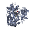
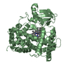
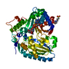
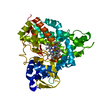
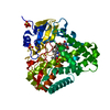
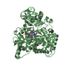
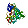
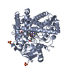
steroid 17-alpha-monooxygenase activity / hormone biosynthetic process / progesterone metabolic process / iron ion binding / heme binding / membrane Similarity search - Function
Monochromator: C(111) / Protocol: SINGLE WAVELENGTH / Monochromatic (M) / Laue (L): M / Scattering type: x-ray
Radiation wavelength
Wavelength: 0.97872 Å / Relative weight: 1
Reflection
Resolution: 2.7→17.08 Å / Num. obs: 22643 / % possible obs: 99 % / Observed criterion σ(I): 5 / Redundancy: 4.7 % / Rmerge(I) obs: 0.074 / Net I/σ(I): 16.4
Reflection shell
Resolution: 2.73→2.81 Å / Redundancy: 4.8 % / Rmerge(I) obs: 0.668 / Mean I/σ(I) obs: 2.2 / % possible all: 98.2
-
Processing
Software
Name
Version
Classification
MD2
diffractometer software from EMBL (with LS-CAT developed extensions)
datacollection
PHASER
phasing
REFMAC
5.8.0073
refinement
HKL-2000
datareduction
HKL-2000
datascaling
Refinement
Method to determine structure: MOLECULAR REPLACEMENT / Resolution: 2.7→17.08 Å / Cor.coef. Fo:Fc: 0.943 / Cor.coef. Fo:Fc free: 0.883 / SU B: 41.57 / SU ML: 0.376 / Cross valid method: THROUGHOUT / ESU R Free: 0.437 / Stereochemistry target values: MAXIMUM LIKELIHOOD / Details: HYDROGENS HAVE BEEN ADDED IN THE RIDING POSITIONS
Rfactor
Num. reflection
% reflection
Selection details
Rfree
0.283
1159
5.1 %
RANDOM
Rwork
0.205
-
-
-
obs
0.209
21459
98.8 %
-
Solvent computation
Ion probe radii: 0.8 Å / Shrinkage radii: 0.8 Å / VDW probe radii: 1.2 Å / Solvent model: MASK
 Movie
Movie Controller


 Open data
 Basic information
 Components
 Keywords
 Function and homology information
 X-RAY DIFFRACTION /
  Authors
 Citation
 Structure visualization
 Downloads & links
 Other downloads
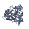


 PDBj
PDBj
 Assembly
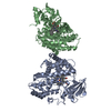

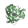

 Mass: 616.487 Da / Num. of mol.: 2 / Source method: obtained synthetically / Formula: C34H32FeN4O4
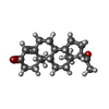
 Mass: 314.462 Da / Num. of mol.: 2 / Source method: obtained synthetically / Formula: C21H30O2
 Mass: 18.015 Da / Num. of mol.: 17 / Source method: isolated from a natural source / Formula: H2O
 Sample preparation
 / Beamline: 21-ID-F / Wavelength: 0.97872
  Processing