Movie
Movie Controller


+ Open data

- Basic information
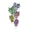
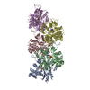
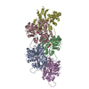
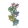
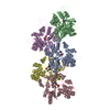
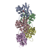
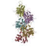
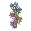
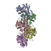
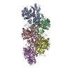
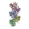
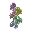
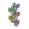
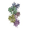
| Entry | Database: PDB / ID: 7bte | |||||||||
|---|---|---|---|---|---|---|---|---|---|---|
| Title | Lifeact-F-actin complex | |||||||||
 Components Components |
| |||||||||
 Keywords Keywords | CONTRACTILE PROTEIN/PROTEIN BINDING / F-actin / ADP-F-actin / CONTRACTILE PROTEIN-PROTEIN BINDING complex | |||||||||
| Function / homology |  Function and homology information Function and homology informationtRNAThr (cytosine32-N3)-methyltransferase / tRNA (cytidine-N3)-methyltransferase activity / Regulation of CDH1 Function / Striated Muscle Contraction / tRNA methylation / mating projection tip / actin filament bundle / striated muscle thin filament / skeletal muscle thin filament assembly / skeletal muscle fiber development ...tRNAThr (cytosine32-N3)-methyltransferase / tRNA (cytidine-N3)-methyltransferase activity / Regulation of CDH1 Function / Striated Muscle Contraction / tRNA methylation / mating projection tip / actin filament bundle / striated muscle thin filament / skeletal muscle thin filament assembly / skeletal muscle fiber development / stress fiber / actin filament / Hydrolases; Acting on acid anhydrides; Acting on acid anhydrides to facilitate cellular and subcellular movement / actin filament binding / actin cytoskeleton / protein-macromolecule adaptor activity / viral translational frameshifting / hydrolase activity / ATP binding / cytoplasm Similarity search - Function | |||||||||
| Biological species |   | |||||||||
| Method | ELECTRON MICROSCOPY / helical reconstruction / cryo EM / Resolution: 4.2 Å | |||||||||
 Authors Authors | Kumari, A. / Ragunath, V.K. / Sirajuddin, M. | |||||||||
| Funding support |  India, 2items India, 2items
| |||||||||
 Citation Citation | Journal: EMBO J / Year: 2020 Title: Structural insights into actin filament recognition by commonly used cellular actin markers. Authors: Archana Kumari / Shubham Kesarwani / Manjunath G Javoor / Kutti R Vinothkumar / Minhajuddin Sirajuddin / Abstract: Cellular studies of filamentous actin (F-actin) processes commonly utilize fluorescent versions of toxins, peptides, and proteins that bind actin. While the choice of these markers has been largely ...Cellular studies of filamentous actin (F-actin) processes commonly utilize fluorescent versions of toxins, peptides, and proteins that bind actin. While the choice of these markers has been largely based on availability and ease, there is a severe dearth of structural data for an informed judgment in employing suitable F-actin markers for a particular requirement. Here, we describe the electron cryomicroscopy structures of phalloidin, lifeAct, and utrophin bound to F-actin, providing a comprehensive high-resolution structural comparison of widely used actin markers and their influence towards F-actin. Our results show that phalloidin binding does not induce specific conformational change and lifeAct specifically recognizes closed D-loop conformation, i.e., ADP-Pi or ADP states of F-actin. The structural models aided designing of minimal utrophin and a shorter lifeAct, which can be utilized as F-actin marker. Together, our study provides a structural perspective, where the binding sites of utrophin and lifeAct overlap with majority of actin-binding proteins and thus offering an invaluable resource for researchers in choosing appropriate actin markers and generating new marker variants. | |||||||||
| History |
|
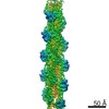
- Structure visualization
| Movie |
Movie viewer |
|---|---|
| Structure viewer | Molecule: MolmilJmol/JSmol |
- Downloads & links
-Download
| PDBx/mmCIF format | 7bte.cif.gz | 344.4 KB | Display | PDBx/mmCIF format |
|---|---|---|---|---|
| PDB format | pdb7bte.ent.gz | 269.7 KB | Display | PDB format |
| PDBx/mmJSON format | 7bte.json.gz | Tree view | PDBx/mmJSON format | |
| Others |  Other downloads Other downloads |
-Validation report
| Arichive directory | https://data.pdbj.org/pub/pdb/validation_reports/bt/7bteftp://data.pdbj.org/pub/pdb/validation_reports/bt/7bte | HTTPS FTP |
|---|
-Related structure data
| Related structure data | 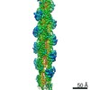 30177MC  6m5gC  7bt7C  7btiC C: citing same article ( M: map data used to model this data |
|---|---|
| Similar structure data |


-Links
 PDBj
PDBj


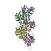
- Assembly
| Deposited unit | 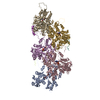
|
|---|---|
| 1 |
|
-Components
| #1: Protein | Mass: 42096.953 Da / Num. of mol.: 5 / Source method: isolated from a natural source / Source: (natural) #2: Protein/peptide | Mass: 1927.243 Da / Num. of mol.: 3 / Source method: obtained synthetically / Source: (synth.) #3: Chemical | ChemComp-MG /   Mass: 24.305 Da / Num. of mol.: 5 / Source method: obtained synthetically / Formula: Mg / Feature type: SUBJECT OF INVESTIGATION Mass: 24.305 Da / Num. of mol.: 5 / Source method: obtained synthetically / Formula: Mg / Feature type: SUBJECT OF INVESTIGATION#4: Chemical | ChemComp-ADP /   Mass: 427.201 Da / Num. of mol.: 5 / Source method: obtained synthetically / Formula: C10H15N5O10P2 / Feature type: SUBJECT OF INVESTIGATION / Comment: ADP, energy-carrying molecule*YM Mass: 427.201 Da / Num. of mol.: 5 / Source method: obtained synthetically / Formula: C10H15N5O10P2 / Feature type: SUBJECT OF INVESTIGATION / Comment: ADP, energy-carrying molecule*YMHas ligand of interest | Y | |
|---|
-Experimental details
-Experiment
| Experiment | Method: ELECTRON MICROSCOPY |
|---|---|
| EM experiment | Aggregation state: FILAMENT / 3D reconstruction method: helical reconstruction |
- Sample preparation
| Component | Name: Filamentous actin in ADP state / Type: COMPLEX / Entity ID: #1-#2 / Source: NATURAL |
|---|---|
| Molecular weight | Experimental value: NO |
| Source (natural) | Organism: |
| Buffer solution | pH: 7.5 Details: 50mM KCl,1mM MgCl2,0.2mM EGTA, 10mM Imidazole buffer pH 7.5 |
| Specimen | Conc.: 0.0002 mg/ml / Embedding applied: NO / Shadowing applied: NO / Staining applied: NO / Vitrification applied: YES Details: 10 mole excess of lifeact was mixed with F-actin and used for sample preparation. |
| Specimen support | Details: No prior coating / Grid material: GOLD / Grid mesh size: 300 divisions/in. / Grid type: Quantifoil R1.2/1.3 |
| Vitrification | Instrument: FEI VITROBOT MARK I / Cryogen name: ETHANE / Humidity: 95 % / Chamber temperature: 293 K / Details: blot for 3.5 seconds |
- Electron microscopy imaging
| Experimental equipment |  Model: Titan Krios / Image courtesy: FEI Company |
|---|---|
| Microscopy | Model: FEI TITAN KRIOS |
| Electron gun | Electron source:  FIELD EMISSION GUN / Accelerating voltage: 300 kV / Illumination mode: OTHER FIELD EMISSION GUN / Accelerating voltage: 300 kV / Illumination mode: OTHER |
| Electron lens | Mode: OTHER / Calibrated magnification: 75000 X / Nominal defocus min: 1500 nm / Cs: 2.7 mm / C2 aperture diameter: 50 µm / Alignment procedure: COMA FREE |
| Specimen holder | Cryogen: NITROGEN / Specimen holder model: FEI TITAN KRIOS AUTOGRID HOLDER / Temperature (max): 120 K / Temperature (min): 100 K |
| Image recording | Average exposure time: 2 sec. / Electron dose: 49.2 e/Å2 / Detector mode: INTEGRATING / Film or detector model: FEI FALCON III (4k x 4k) / Num. of grids imaged: 1 / Num. of real images: 529 / Details: 30 eV |
- Processing
| Software |
| |||||||||||||||||||||||||||||||||||||||||||||
|---|---|---|---|---|---|---|---|---|---|---|---|---|---|---|---|---|---|---|---|---|---|---|---|---|---|---|---|---|---|---|---|---|---|---|---|---|---|---|---|---|---|---|---|---|---|---|
| EM software |
| |||||||||||||||||||||||||||||||||||||||||||||
| CTF correction | Details: GCTF for CTF correction / Type: NONE | |||||||||||||||||||||||||||||||||||||||||||||
| Helical symmerty | Angular rotation/subunit: -166.9 ° / Axial rise/subunit: 27.44 Å / Axial symmetry: C1 | |||||||||||||||||||||||||||||||||||||||||||||
| Particle selection | Num. of particles selected: 111074 | |||||||||||||||||||||||||||||||||||||||||||||
| 3D reconstruction | Resolution: 4.2 Å / Resolution method: FSC 0.143 CUT-OFF / Num. of particles: 297584 / Num. of class averages: 100 / Symmetry type: HELICAL | |||||||||||||||||||||||||||||||||||||||||||||
| Atomic model building | B value: 179 / Protocol: RIGID BODY FIT / Space: REAL | |||||||||||||||||||||||||||||||||||||||||||||
| Atomic model building | PDB-ID: 5ONV Pdb chain-ID: A / Accession code: 5ONV / Pdb chain residue range: 6-373 / Source name: PDB / Type: experimental model | |||||||||||||||||||||||||||||||||||||||||||||
| Refinement | Stereochemistry target values: GeoStd + Monomer Library + CDL v1.2 | |||||||||||||||||||||||||||||||||||||||||||||
| Refine LS restraints |
|