 Movie
Movie Controller

[English] 日本語
 Yorodumi
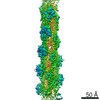
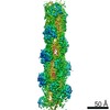
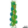
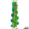
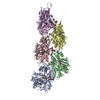
- PDB-5ood: Cryo-EM structure of jasplakinolide-stabilized F-actin in complex... -
+ Open data

- Basic information
| Entry | Database: PDB / ID: 5ood | ||||||||||||||||||||||||||||||||||||||||||||||||||||||||||||
|---|---|---|---|---|---|---|---|---|---|---|---|---|---|---|---|---|---|---|---|---|---|---|---|---|---|---|---|---|---|---|---|---|---|---|---|---|---|---|---|---|---|---|---|---|---|---|---|---|---|---|---|---|---|---|---|---|---|---|---|---|---|
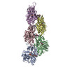
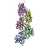
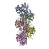
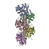
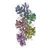
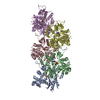
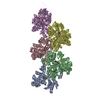
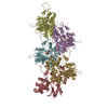
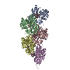
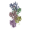
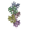
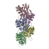
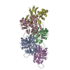
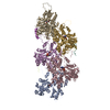
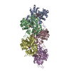
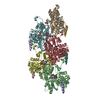
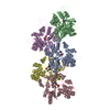
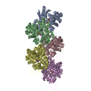
| Title | Cryo-EM structure of jasplakinolide-stabilized F-actin in complex with ADP-Pi | ||||||||||||||||||||||||||||||||||||||||||||||||||||||||||||
 Components Components | Actin, alpha skeletal muscle | ||||||||||||||||||||||||||||||||||||||||||||||||||||||||||||
 Keywords Keywords | STRUCTURAL PROTEIN / Cytoskeleton / nucleotide states / filament stability / cell migration | ||||||||||||||||||||||||||||||||||||||||||||||||||||||||||||
| Function / homology |  Function and homology information Function and homology informationcytoskeletal motor activator activity / myosin heavy chain binding / tropomyosin binding / actin filament bundle / troponin I binding / filamentous actin / mesenchyme migration / skeletal muscle myofibril / actin filament bundle assembly / striated muscle thin filament ...cytoskeletal motor activator activity / myosin heavy chain binding / tropomyosin binding / actin filament bundle / troponin I binding / filamentous actin / mesenchyme migration / skeletal muscle myofibril / actin filament bundle assembly / striated muscle thin filament / skeletal muscle thin filament assembly / actin monomer binding / skeletal muscle fiber development / stress fiber / titin binding / actin filament polymerization / filopodium / actin filament / Hydrolases; Acting on acid anhydrides; Acting on acid anhydrides to facilitate cellular and subcellular movement / calcium-dependent protein binding / lamellipodium / cell body / protein domain specific binding / hydrolase activity / calcium ion binding / positive regulation of gene expression / magnesium ion binding / ATP binding / identical protein binding / cytoplasm Similarity search - Function | ||||||||||||||||||||||||||||||||||||||||||||||||||||||||||||
| Biological species |  | ||||||||||||||||||||||||||||||||||||||||||||||||||||||||||||
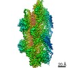
| Method | ELECTRON MICROSCOPY / single particle reconstruction / cryo EM / Resolution: 3.7 Å | ||||||||||||||||||||||||||||||||||||||||||||||||||||||||||||
 Authors Authors | Merino, F. / Pospich, S. / Funk, J. / Kuellmer, F. / Arndt, H.-D. / Bieling, P. / Raunser, S. | ||||||||||||||||||||||||||||||||||||||||||||||||||||||||||||
| Funding support |  Germany, 4items Germany, 4items
| ||||||||||||||||||||||||||||||||||||||||||||||||||||||||||||
 Citation Citation | Journal: Nat Struct Mol Biol / Year: 2018 Title: Structural transitions of F-actin upon ATP hydrolysis at near-atomic resolution revealed by cryo-EM. Authors: Felipe Merino / Sabrina Pospich / Johanna Funk / Thorsten Wagner / Florian Küllmer / Hans-Dieter Arndt / Peter Bieling / Stefan Raunser / Abstract: The function of actin is coupled to the nucleotide bound to its active site. ATP hydrolysis is activated during polymerization; a delay between hydrolysis and inorganic phosphate (P) release results ...The function of actin is coupled to the nucleotide bound to its active site. ATP hydrolysis is activated during polymerization; a delay between hydrolysis and inorganic phosphate (P) release results in a gradient of ATP, ADP-P and ADP along actin filaments (F-actin). Actin-binding proteins can recognize F-actin's nucleotide state, using it as a local 'age' tag. The underlying mechanism is complex and poorly understood. Here we report six high-resolution cryo-EM structures of F-actin from rabbit skeletal muscle in different nucleotide states. The structures reveal that actin polymerization repositions the proposed catalytic base, His161, closer to the γ-phosphate. Nucleotide hydrolysis and P release modulate the conformational ensemble at the periphery of the filament, thus resulting in open and closed states, which can be sensed by coronin-1B. The drug-like toxin jasplakinolide locks F-actin in an open state. Our results demonstrate in detail how ATP hydrolysis links to F-actin's conformational dynamics and protein interaction. | ||||||||||||||||||||||||||||||||||||||||||||||||||||||||||||
| History |
|
- Structure visualization
| Movie |
Movie viewer |
|---|---|
| Structure viewer | Molecule: MolmilJmol/JSmol |
- Downloads & links
-Download
| PDBx/mmCIF format | 5ood.cif.gz | 371.5 KB | Display | PDBx/mmCIF format |
|---|---|---|---|---|
| PDB format | pdb5ood.ent.gz | 303.4 KB | Display | PDB format |
| PDBx/mmJSON format | 5ood.json.gz | Tree view | PDBx/mmJSON format | |
| Others |  Other downloads Other downloads |
-Validation report
| Arichive directory | https://data.pdbj.org/pub/pdb/validation_reports/oo/5oodftp://data.pdbj.org/pub/pdb/validation_reports/oo/5ood | HTTPS FTP |
|---|
-Related structure data
| Related structure data | 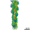 3837MC 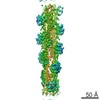 3835C  3836C 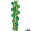 3838C  3839C 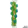 4259C 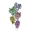 5onvC  5oocC  5ooeC 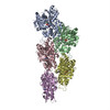 5oofC 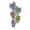 6fhlC C: citing same article ( M: map data used to model this data |
|---|---|
| Similar structure data |


-Links
 PDBj
PDBj


- Assembly
| Deposited unit | 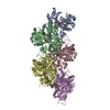
|
|---|---|
| 1 |
|
-Components
| #1: Protein | Mass: 41875.633 Da / Num. of mol.: 5 / Source method: isolated from a natural source / Source: (natural) #2: Chemical | ChemComp-ADP /   Mass: 427.201 Da / Num. of mol.: 5 / Source method: obtained synthetically / Formula: C10H15N5O10P2 / Comment: ADP, energy-carrying molecule*YM Mass: 427.201 Da / Num. of mol.: 5 / Source method: obtained synthetically / Formula: C10H15N5O10P2 / Comment: ADP, energy-carrying molecule*YM#3: Chemical | ChemComp-PO4 /   Mass: 94.971 Da / Num. of mol.: 5 / Source method: obtained synthetically / Formula: PO4 Mass: 94.971 Da / Num. of mol.: 5 / Source method: obtained synthetically / Formula: PO4#4: Chemical | ChemComp-9ZK / (   Mass: 673.841 Da / Num. of mol.: 5 / Source method: obtained synthetically / Formula: C38H51N5O6 Mass: 673.841 Da / Num. of mol.: 5 / Source method: obtained synthetically / Formula: C38H51N5O6#5: Chemical | ChemComp-MG /   Mass: 24.305 Da / Num. of mol.: 5 / Source method: obtained synthetically / Formula: Mg Mass: 24.305 Da / Num. of mol.: 5 / Source method: obtained synthetically / Formula: MgHas protein modification | Y | |
|---|
-Experimental details
-Experiment
| Experiment | Method: ELECTRON MICROSCOPY |
|---|---|
| EM experiment | Aggregation state: FILAMENT / 3D reconstruction method: single particle reconstruction |
| Crystal symmetry | ∠γ: 90 ° / A: 1 Å / B: 1 Å / C: 1 Å / Space group name H-M: P1 |
- Sample preparation
| Component | Name: Filamentous jasplakinolide-stabilized alpha actin in complex with ADP-Pi Type: COMPLEX / Entity ID: #1 / Source: NATURAL | |||||||||||||||||||||||||||||||||||
|---|---|---|---|---|---|---|---|---|---|---|---|---|---|---|---|---|---|---|---|---|---|---|---|---|---|---|---|---|---|---|---|---|---|---|---|---|
| Molecular weight | Experimental value: NO | |||||||||||||||||||||||||||||||||||
| Source (natural) | Organism: | |||||||||||||||||||||||||||||||||||
| Buffer solution | pH: 7.5 Details: 5 mM Tris pH 7.5, 2 mM NaN3, 1 mM DTT, 100 mM KCl and 2 mM MgCl2, 9.0 %(v/v) DMSO | |||||||||||||||||||||||||||||||||||
| Buffer component |
| |||||||||||||||||||||||||||||||||||
| Specimen | Embedding applied: NO / Shadowing applied: NO / Staining applied: NO / Vitrification applied: YES / Details: Rise 27.39 A, Twist -166.41 degrees | |||||||||||||||||||||||||||||||||||
| Specimen support | Grid material: COPPER / Grid mesh size: 300 divisions/in. / Grid type: Quantifoil R2/1 | |||||||||||||||||||||||||||||||||||
| Vitrification | Instrument: GATAN CRYOPLUNGE 3 / Cryogen name: ETHANE / Humidity: 90 % / Chamber temperature: 298 K Details: Manual backside blotting using Whatman filter paper No.5. |
- Electron microscopy imaging
| Experimental equipment |  Model: Titan Krios / Image courtesy: FEI Company |
|---|---|
| Microscopy | Model: FEI TITAN KRIOS / Details: Cs-corrected microscope |
| Electron gun | Electron source:  FIELD EMISSION GUN / Accelerating voltage: 300 kV / Illumination mode: SPOT SCAN FIELD EMISSION GUN / Accelerating voltage: 300 kV / Illumination mode: SPOT SCAN |
| Electron lens | Mode: BRIGHT FIELD / Alignment procedure: COMA FREE |
| Specimen holder | Cryogen: NITROGEN |
| Image recording | Average exposure time: 15 sec. / Electron dose: 78 e/Å2 / Detector mode: SUPER-RESOLUTION / Film or detector model: GATAN K2 SUMMIT (4k x 4k) / Num. of real images: 724 / Details: Images were binned twice during motion correction. |
| EM imaging optics | Energyfilter name: GIF Quantum ER / Spherical aberration corrector: Cs-corrected microscope |
| Image scans | Movie frames/image: 75 / Used frames/image: 1-15 |
- Processing
| EM software |
| |||||||||||||||||||||||||||||||||||||||||||||||||||||||
|---|---|---|---|---|---|---|---|---|---|---|---|---|---|---|---|---|---|---|---|---|---|---|---|---|---|---|---|---|---|---|---|---|---|---|---|---|---|---|---|---|---|---|---|---|---|---|---|---|---|---|---|---|---|---|---|---|
| Crystal symmetry | ∠γ: 90 ° / A: 1 Å / B: 1 Å / C: 1 Å / Space group name H-M: P1 | |||||||||||||||||||||||||||||||||||||||||||||||||||||||
| CTF correction | Type: PHASE FLIPPING AND AMPLITUDE CORRECTION | |||||||||||||||||||||||||||||||||||||||||||||||||||||||
| Particle selection | Num. of particles selected: 154223 | |||||||||||||||||||||||||||||||||||||||||||||||||||||||
| Symmetry | Point symmetry: C1 (asymmetric) | |||||||||||||||||||||||||||||||||||||||||||||||||||||||
| 3D reconstruction | Resolution: 3.7 Å / Resolution method: FSC 0.143 CUT-OFF / Num. of particles: 123716 / Algorithm: BACK PROJECTION / Symmetry type: POINT | |||||||||||||||||||||||||||||||||||||||||||||||||||||||
| Atomic model building | Protocol: OTHER / Space: REAL Details: Rosetta iterative refinement was combined with MDFF. |
