 Movie
Movie Controller


+ Open data

- Basic information
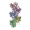
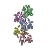
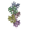
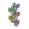
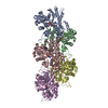
| Entry | Database: PDB / ID: 3j8i | ||||||
|---|---|---|---|---|---|---|---|
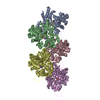
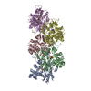
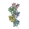
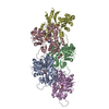
| Title | Near-Atomic Resolution for One State of F-Actin | ||||||
 Components Components | Actin, alpha skeletal muscle | ||||||
 Keywords Keywords | STRUCTURAL PROTEIN / helical polymer / F-actin | ||||||
| Function / homology |  Function and homology information Function and homology informationcytoskeletal motor activator activity / myosin heavy chain binding / tropomyosin binding / actin filament bundle / troponin I binding / filamentous actin / mesenchyme migration / skeletal muscle myofibril / actin filament bundle assembly / striated muscle thin filament ...cytoskeletal motor activator activity / myosin heavy chain binding / tropomyosin binding / actin filament bundle / troponin I binding / filamentous actin / mesenchyme migration / skeletal muscle myofibril / actin filament bundle assembly / striated muscle thin filament / skeletal muscle thin filament assembly / actin monomer binding / skeletal muscle fiber development / stress fiber / titin binding / actin filament polymerization / filopodium / actin filament / Hydrolases; Acting on acid anhydrides; Acting on acid anhydrides to facilitate cellular and subcellular movement / calcium-dependent protein binding / lamellipodium / cell body / protein domain specific binding / hydrolase activity / calcium ion binding / positive regulation of gene expression / magnesium ion binding / ATP binding / identical protein binding / cytoplasm Similarity search - Function | ||||||
| Biological species |  | ||||||
| Method | ELECTRON MICROSCOPY / helical reconstruction / cryo EM / Resolution: 4.7 Å | ||||||
 Authors Authors | Galkin, V.E. | ||||||
 Citation Citation | Journal: Structure / Year: 2015 Title: Near-atomic resolution for one state of F-actin. Authors: Vitold E Galkin / Albina Orlova / Matthijn R Vos / Gunnar F Schröder / Edward H Egelman /    Abstract: Actin functions as a helical polymer, F-actin, but attempts to build an atomic model for this filament have been hampered by the fact that the filament cannot be crystallized and by structural ...Actin functions as a helical polymer, F-actin, but attempts to build an atomic model for this filament have been hampered by the fact that the filament cannot be crystallized and by structural heterogeneity. We have used a direct electron detector, cryo-electron microscopy, and the forces imposed on actin filaments in thin films to reconstruct one state of the filament at 4.7 Å resolution, which allows for building a reliable pseudo-atomic model of F-actin. We also report a different state of the filament where actin protomers adopt a conformation observed in the crystal structure of the G-actin-profilin complex with an open ATP-binding cleft. Comparison of the two structural states provides insights into ATP-hydrolysis and filament dynamics. The atomic model provides a framework for understanding why every buried residue in actin has been under intense selective pressure. | ||||||
| History |
|
- Structure visualization
| Movie |
Movie viewer |
|---|---|
| Structure viewer | Molecule: MolmilJmol/JSmol |
- Downloads & links
-Download
| PDBx/mmCIF format | 3j8i.cif.gz | 335.9 KB | Display | PDBx/mmCIF format |
|---|---|---|---|---|
| PDB format | pdb3j8i.ent.gz | 278 KB | Display | PDB format |
| PDBx/mmJSON format | 3j8i.json.gz | Tree view | PDBx/mmJSON format | |
| Others |  Other downloads Other downloads |
-Validation report
| Arichive directory | https://data.pdbj.org/pub/pdb/validation_reports/j8/3j8iftp://data.pdbj.org/pub/pdb/validation_reports/j8/3j8i | HTTPS FTP |
|---|
-Related structure data
| Related structure data | 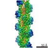 6179MC 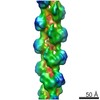 6180C 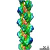 6181C 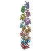 3j8jC 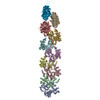 3j8kC M: map data used to model this data C: citing same article ( |
|---|---|
| Similar structure data |
-Links
 PDBj
PDBj


- Assembly
| Deposited unit | 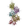
|
|---|---|
| 1 |
|
| Symmetry | Helical symmetry: (Circular symmetry: 1 / Dyad axis: no / N subunits divisor: 1 / Num. of operations: 5 / Rise per n subunits: 27.6 Å / Rotation per n subunits: -166.7 °) |
| Details | Actin forms a helical filament of indeterminate length. The designation "pentameric" in REMARK 350 is an artifact of the PDB format and can be disregarded. |
-Components
| #1: Protein | Mass: 42096.953 Da / Num. of mol.: 5 / Source method: isolated from a natural source / Source: (natural) #2: Chemical | ChemComp-ADP /   Mass: 427.201 Da / Num. of mol.: 5 / Source method: obtained synthetically / Formula: C10H15N5O10P2 / Comment: ADP, energy-carrying molecule*YM Mass: 427.201 Da / Num. of mol.: 5 / Source method: obtained synthetically / Formula: C10H15N5O10P2 / Comment: ADP, energy-carrying molecule*YM#3: Chemical | ChemComp-MG /   Mass: 24.305 Da / Num. of mol.: 5 / Source method: obtained synthetically / Formula: Mg Mass: 24.305 Da / Num. of mol.: 5 / Source method: obtained synthetically / Formula: Mg |
|---|
-Experimental details
-Experiment
| Experiment | Method: ELECTRON MICROSCOPY |
|---|---|
| EM experiment | Aggregation state: FILAMENT / 3D reconstruction method: helical reconstruction |
- Sample preparation
| Component | Name: Skeletal muscle actin / Type: COMPLEX / Details: polymer |
|---|---|
| Specimen | Embedding applied: NO / Shadowing applied: NO / Staining applied: NO / Vitrification applied: YES |
| Vitrification | Instrument: FEI VITROBOT MARK IV / Cryogen name: ETHANE / Humidity: 90 % / Details: Plunged into liquid ethane (FEI VITROBOT MARK IV) |
- Electron microscopy imaging
| Experimental equipment |  Model: Titan Krios / Image courtesy: FEI Company |
|---|---|
| Microscopy | Model: FEI TITAN KRIOS / Date: Oct 9, 2014 |
| Electron gun | Electron source:  FIELD EMISSION GUN / Accelerating voltage: 300 kV / Illumination mode: FLOOD BEAM FIELD EMISSION GUN / Accelerating voltage: 300 kV / Illumination mode: FLOOD BEAM |
| Electron lens | Mode: BRIGHT FIELD / Nominal defocus max: 3000 nm / Nominal defocus min: 500 nm / Cs: 2.7 mm |
| Specimen holder | Specimen holder model: FEI TITAN KRIOS AUTOGRID HOLDER |
| Image recording | Film or detector model: FEI FALCON II (4k x 4k) |
| Image scans | Num. digital images: 850 |
- Processing
| EM software |
| ||||||||||||
|---|---|---|---|---|---|---|---|---|---|---|---|---|---|
| Helical symmerty | Angular rotation/subunit: 166.7 ° / Axial rise/subunit: 27.6 Å / Axial symmetry: C1 | ||||||||||||
| 3D reconstruction | Resolution: 4.7 Å / Resolution method: FSC / Nominal pixel size: 1.05 Å / Actual pixel size: 1.05 Å / Details: (Helical Details: IHRSR) / Symmetry type: HELICAL | ||||||||||||
| Refinement step | Cycle: LAST
|
