Movie
Movie Controller


+ Open data

- Basic information
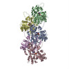
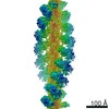
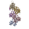
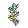
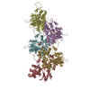
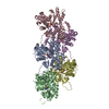
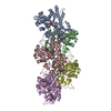
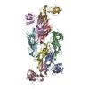
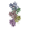
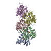
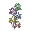
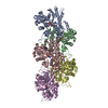
| Entry | Database: PDB / ID: 6tu4 | |||||||||
|---|---|---|---|---|---|---|---|---|---|---|
| Title | Structure of Plasmodium Actin1 filament | |||||||||
 Components Components | Actin-1 | |||||||||
 Keywords Keywords | MOTOR PROTEIN / malaria / Plasmodium falciparum / myosin / unconventional / filament | |||||||||
| Function / homology |  Function and homology information Function and homology informationplastid inheritance / schizogony / symbiont-mediated actin polymerization-dependent cell-to-cell migration in host / Platelet degranulation / entry into host cell by a symbiont-containing vacuole / cytoskeleton organization / actin filament / structural constituent of cytoskeleton / Hydrolases; Acting on acid anhydrides; Acting on acid anhydrides to facilitate cellular and subcellular movement / actin cytoskeleton ...plastid inheritance / schizogony / symbiont-mediated actin polymerization-dependent cell-to-cell migration in host / Platelet degranulation / entry into host cell by a symbiont-containing vacuole / cytoskeleton organization / actin filament / structural constituent of cytoskeleton / Hydrolases; Acting on acid anhydrides; Acting on acid anhydrides to facilitate cellular and subcellular movement / actin cytoskeleton / ATP hydrolysis activity / ATP binding / nucleus / cytoplasm Similarity search - Function | |||||||||
| Biological species |  | |||||||||
| Method | ELECTRON MICROSCOPY / helical reconstruction / cryo EM / Resolution: 2.6 Å | |||||||||
 Authors Authors | Vahokoski, J. / Calder, L.J. / Lopez, A.J. / Rosenthal, P.B. / Kursula, I. | |||||||||
| Funding support |  Norway, Norway,  United Kingdom, 2items United Kingdom, 2items
| |||||||||
 Citation Citation | Journal: PLoS Pathog / Year: 2022 Title: High-resolution structures of malaria parasite actomyosin and actin filaments. Authors: Juha Vahokoski / Lesley J Calder / Andrea J Lopez / Justin E Molloy / Inari Kursula / Peter B Rosenthal /  Abstract: Malaria is responsible for half a million deaths annually and poses a huge economic burden on the developing world. The mosquito-borne parasites (Plasmodium spp.) that cause the disease depend upon ...Malaria is responsible for half a million deaths annually and poses a huge economic burden on the developing world. The mosquito-borne parasites (Plasmodium spp.) that cause the disease depend upon an unconventional actomyosin motor for both gliding motility and host cell invasion. The motor system, often referred to as the glideosome complex, remains to be understood in molecular terms and is an attractive target for new drugs that might block the infection pathway. Here, we present the high-resolution structure of the actomyosin motor complex from Plasmodium falciparum. The complex includes the malaria parasite actin filament (PfAct1) complexed with the class XIV myosin motor (PfMyoA) and its two associated light-chains. The high-resolution core structure reveals the PfAct1:PfMyoA interface in atomic detail, while at lower-resolution, we visualize the PfMyoA light-chain binding region, including the essential light chain (PfELC) and the myosin tail interacting protein (PfMTIP). Finally, we report a bare PfAct1 filament structure at improved resolution. | |||||||||
| History |
|
- Structure visualization
| Movie |
Movie viewer |
|---|---|
| Structure viewer | Molecule: MolmilJmol/JSmol |
- Downloads & links
-Download
| PDBx/mmCIF format | 6tu4.cif.gz | 353.9 KB | Display | PDBx/mmCIF format |
|---|---|---|---|---|
| PDB format | pdb6tu4.ent.gz | 284.9 KB | Display | PDB format |
| PDBx/mmJSON format | 6tu4.json.gz | Tree view | PDBx/mmJSON format | |
| Others |  Other downloads Other downloads |
-Validation report
| Arichive directory | https://data.pdbj.org/pub/pdb/validation_reports/tu/6tu4ftp://data.pdbj.org/pub/pdb/validation_reports/tu/6tu4 | HTTPS FTP |
|---|
-Related structure data
| Related structure data | 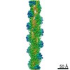 10587MC  6tu7C M: map data used to model this data C: citing same article ( |
|---|---|
| Similar structure data |
-Links
 PDBj
PDBj


- Assembly
| Deposited unit | 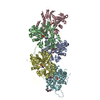
|
|---|---|
| 1 |
|
-Components
| #1: Protein | Mass: 42047.676 Da / Num. of mol.: 5 Source method: isolated from a genetically manipulated source Details: codon optimised gene / Source: (gene. exp.)   Spodoptera frugiperda (fall armyworm) / References: UniProt: Q8I4X0 Spodoptera frugiperda (fall armyworm) / References: UniProt: Q8I4X0#2: Chemical | ChemComp-MG /   Mass: 24.305 Da / Num. of mol.: 5 Mass: 24.305 Da / Num. of mol.: 5Source method: isolated from a genetically manipulated source Formula: Mg #3: Chemical | ChemComp-9UE /   Mass: 709.670 Da / Num. of mol.: 5 / Source method: obtained synthetically / Formula: C36H45BrN4O6 Mass: 709.670 Da / Num. of mol.: 5 / Source method: obtained synthetically / Formula: C36H45BrN4O6#4: Chemical | ChemComp-ADP /   Mass: 427.201 Da / Num. of mol.: 5 / Source method: obtained synthetically / Formula: C10H15N5O10P2 / Feature type: SUBJECT OF INVESTIGATION / Comment: ADP, energy-carrying molecule*YM Mass: 427.201 Da / Num. of mol.: 5 / Source method: obtained synthetically / Formula: C10H15N5O10P2 / Feature type: SUBJECT OF INVESTIGATION / Comment: ADP, energy-carrying molecule*YM#5: Water | ChemComp-HOH / |  Mass: 18.015 Da / Num. of mol.: 140 / Source method: isolated from a natural source / Formula: H2O Mass: 18.015 Da / Num. of mol.: 140 / Source method: isolated from a natural source / Formula: H2OHas ligand of interest | Y | |
|---|
-Experimental details
-Experiment
| Experiment | Method: ELECTRON MICROSCOPY |
|---|---|
| EM experiment | Aggregation state: FILAMENT / 3D reconstruction method: helical reconstruction |
- Sample preparation
| Component | Name: PfAct1 filament / Type: COMPLEX / Entity ID: #1 / Source: RECOMBINANT |
|---|---|
| Molecular weight | Experimental value: NO |
| Source (natural) | Organism: |
| Source (recombinant) | Organism: Spodoptera frugiperda (fall armyworm) / Cell: Sf21 |
| Buffer solution | pH: 7.5 |
| Specimen | Embedding applied: NO / Shadowing applied: NO / Staining applied: NO / Vitrification applied: YES |
| Vitrification | Cryogen name: ETHANE |
- Electron microscopy imaging
| Experimental equipment |  Model: Titan Krios / Image courtesy: FEI Company |
|---|---|
| Microscopy | Model: FEI TITAN KRIOS |
| Electron gun | Electron source:  FIELD EMISSION GUN / Accelerating voltage: 300 kV / Illumination mode: SPOT SCAN FIELD EMISSION GUN / Accelerating voltage: 300 kV / Illumination mode: SPOT SCAN |
| Electron lens | Mode: BRIGHT FIELD |
| Image recording | Electron dose: 51.52 e/Å2 / Detector mode: COUNTING / Film or detector model: FEI FALCON III (4k x 4k) |
- Processing
| Software |
| ||||||||||||||||||||||||
|---|---|---|---|---|---|---|---|---|---|---|---|---|---|---|---|---|---|---|---|---|---|---|---|---|---|
| EM software |
| ||||||||||||||||||||||||
| CTF correction | Type: PHASE FLIPPING ONLY | ||||||||||||||||||||||||
| Helical symmerty | Angular rotation/subunit: -167.652 ° / Axial rise/subunit: 28.3692 Å / Axial symmetry: C1 | ||||||||||||||||||||||||
| 3D reconstruction | Resolution: 2.6 Å / Resolution method: FSC 0.143 CUT-OFF / Num. of particles: 305480 / Symmetry type: HELICAL | ||||||||||||||||||||||||
| Atomic model building | Space: REAL | ||||||||||||||||||||||||
| Atomic model building | PDB-ID: 5OGW Pdb chain-ID: A / Accession code: 5OGW / Source name: PDB / Type: experimental model | ||||||||||||||||||||||||
| Refinement | Cross valid method: NONE Stereochemistry target values: GeoStd + Monomer Library + CDL v1.2 | ||||||||||||||||||||||||
| Displacement parameters | Biso mean: 37.12 Å2 | ||||||||||||||||||||||||
| Refine LS restraints |
|