 Movie
Movie Controller

[English] 日本語
 Yorodumi
- PDB-7brl: Atrial Natriuretic Peptide Receptor complexed with Deletion mutan... -
+ Open data

- Basic information
| Entry | Database: PDB / ID: 7brl | |||||||||
|---|---|---|---|---|---|---|---|---|---|---|
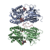
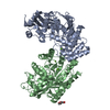
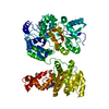
| Title | Atrial Natriuretic Peptide Receptor complexed with Deletion mutant of rat Atrial Natriuretic Peptide[4-17,23] | |||||||||
 Components Components |
| |||||||||
 Keywords Keywords | MEMBRANE PROTEIN / RECEPTOR-HORMONE COMPLEX / NATRIURETIC PEPTIDE / NATRIURETIC PEPTIDE RECEPTOR / GUANYLYL-CYCLASE-COUPLED RECEPTOR / SIGNAL TRANSMISSION / ROTATION MECHANISM / SIGNALING PROTEIN | |||||||||
| Function / homology |  Function and homology information Function and homology informationPhysiological factors / negative regulation of collecting lymphatic vessel constriction / ANPR-A receptor complex / natriuretic peptide receptor activity / cGMP metabolic process / : / neuropeptide receptor binding / mast cell granule / response to 3-methylcholanthrene / cell growth involved in cardiac muscle cell development ...Physiological factors / negative regulation of collecting lymphatic vessel constriction / ANPR-A receptor complex / natriuretic peptide receptor activity / cGMP metabolic process / : / neuropeptide receptor binding / mast cell granule / response to 3-methylcholanthrene / cell growth involved in cardiac muscle cell development / peptide receptor activity / regulation of body fluid levels / positive regulation of potassium ion export across plasma membrane / guanylate cyclase / cGMP biosynthetic process / receptor guanylyl cyclase signaling pathway / guanylate cyclase activity / negative regulation of JUN kinase activity / synaptic signaling via neuropeptide / regulation of atrial cardiac muscle cell membrane repolarization / multicellular organismal-level water homeostasis / cardiac muscle hypertrophy in response to stress / sodium ion export across plasma membrane / : / neuropeptide hormone activity / hormone receptor binding / regulation of vascular permeability / glycinergic synapse / negative regulation of systemic arterial blood pressure / regulation of calcium ion transmembrane transport via high voltage-gated calcium channel / dopamine metabolic process / hormone binding / cellular response to angiotensin / peptide hormone binding / brush border / negative regulation of epidermal growth factor receptor signaling pathway / positive regulation of heart rate / negative regulation of blood pressure / cell projection / positive regulation of cardiac muscle contraction / blood vessel diameter maintenance / cellular response to glucose stimulus / negative regulation of smooth muscle cell proliferation / female pregnancy / negative regulation of canonical Wnt signaling pathway / cellular response to mechanical stimulus / neuropeptide signaling pathway / hormone activity / response to insulin / negative regulation of cell growth / regulation of blood pressure / vasodilation / cellular response to hydrogen peroxide / heart development / protein folding / perikaryon / response to hypoxia / protein kinase activity / cell surface receptor signaling pathway / signaling receptor complex / intracellular signal transduction / signaling receptor binding / GTP binding / perinuclear region of cytoplasm / protein-containing complex / : / ATP binding / plasma membrane / cytoplasm Similarity search - Function | |||||||||
| Biological species |  | |||||||||
| Method |  X-RAY DIFFRACTION / SYNCHROTRON / MOLECULAR REPLACEMENT / Resolution: 3.2 Å X-RAY DIFFRACTION / SYNCHROTRON / MOLECULAR REPLACEMENT / Resolution: 3.2 Å | |||||||||
 Authors Authors | Ogawa, H. | |||||||||
| Funding support |  Japan, 2items Japan, 2items
| |||||||||
 Citation Citation | Journal: To Be Published Title: Structural insight into hormone-recognition and transmembrane signaling by the atrial natriuretic peptide receptor Authors: Ogawa, H. | |||||||||
| History |
|
- Structure visualization
| Structure viewer | Molecule: MolmilJmol/JSmol |
|---|
- Downloads & links
-Download
| PDBx/mmCIF format | 7brl.cif.gz | 367.5 KB | Display | PDBx/mmCIF format |
|---|---|---|---|---|
| PDB format | pdb7brl.ent.gz | 306.2 KB | Display | PDB format |
| PDBx/mmJSON format | 7brl.json.gz | Tree view | PDBx/mmJSON format | |
| Others |  Other downloads Other downloads |
-Validation report
| Arichive directory | https://data.pdbj.org/pub/pdb/validation_reports/br/7brlftp://data.pdbj.org/pub/pdb/validation_reports/br/7brl | HTTPS FTP |
|---|
-Related structure data
| Related structure data |  7brgC  7brhC 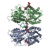 7briC 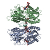 7brjC 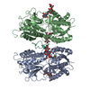 7brkC 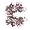 1dp4S S: Starting model for refinement C: citing same article ( |
|---|---|
| Similar structure data |


-Links
 PDBj
PDBj


- Assembly
| Deposited unit | 
| ||||||||
|---|---|---|---|---|---|---|---|---|---|
| 1 |
| ||||||||
| Unit cell |
|
-Components
| #1: Protein | Mass: 48434.828 Da / Num. of mol.: 2 Source method: isolated from a genetically manipulated source Source: (gene. exp.) #2: Protein/peptide | | Mass: 1600.848 Da / Num. of mol.: 1 / Mutation: deletions of residues 1-3, 18-22 and 24-28 / Source method: obtained synthetically / Source: (synth.) #3: Polysaccharide | Source method: isolated from a genetically manipulated source #4: Polysaccharide | Source method: isolated from a genetically manipulated source #5: Chemical |   Mass: 35.453 Da / Num. of mol.: 2 / Source method: obtained synthetically / Formula: Cl / Feature type: SUBJECT OF INVESTIGATION Mass: 35.453 Da / Num. of mol.: 2 / Source method: obtained synthetically / Formula: Cl / Feature type: SUBJECT OF INVESTIGATIONHas ligand of interest | Y | Has protein modification | Y | |
|---|
-Experimental details
-Experiment
| Experiment | Method: X-RAY DIFFRACTION / Number of used crystals: 1 |
|---|
- Sample preparation
| Crystal | Density Matthews: 3.99 Å3/Da / Density % sol: 69.14 % |
|---|---|
| Crystal grow | Temperature: 300 K / Method: vapor diffusion, hanging drop / Details: Sodium malonate, MES / PH range: 6.3 - 6.6 |
-Data collection
| Diffraction | Mean temperature: 100 K / Serial crystal experiment: N |
|---|---|
| Diffraction source | Source: SYNCHROTRON / Site: SPring-8 / Beamline: BL41XU / Wavelength: 0.9 Å |
| Detector | Type: RAYONIX MX225HE / Detector: CCD / Date: Aug 28, 2015 |
| Radiation | Protocol: SINGLE WAVELENGTH / Monochromatic (M) / Laue (L): M / Scattering type: x-ray |
| Radiation wavelength | Wavelength: 0.9 Å / Relative weight: 1 |
| Reflection | Resolution: 3.2→50 Å / Num. obs: 24713 / % possible obs: 97.4 % / Redundancy: 23.1 % / CC1/2: 0.99 / Rmerge(I) obs: 0.048 / Net I/σ(I): 27.6 |
| Reflection shell | Resolution: 3.2→3.29 Å / Rmerge(I) obs: 0.373 / Mean I/σ(I) obs: 3.5 / Num. unique obs: 2096 / CC1/2: 0.42 |
- Processing
| Software |
| |||||||||||||||||||||||||||||||||||||||||||||||||||||||||||||||||||||||||||||||||||||||||||||||||||||||||||||||||||||||||||||||||||||||||||||||||||||||||||||||||||||||||||||||||||||||||||||||||||||||||||||||||||||||||||||||||
|---|---|---|---|---|---|---|---|---|---|---|---|---|---|---|---|---|---|---|---|---|---|---|---|---|---|---|---|---|---|---|---|---|---|---|---|---|---|---|---|---|---|---|---|---|---|---|---|---|---|---|---|---|---|---|---|---|---|---|---|---|---|---|---|---|---|---|---|---|---|---|---|---|---|---|---|---|---|---|---|---|---|---|---|---|---|---|---|---|---|---|---|---|---|---|---|---|---|---|---|---|---|---|---|---|---|---|---|---|---|---|---|---|---|---|---|---|---|---|---|---|---|---|---|---|---|---|---|---|---|---|---|---|---|---|---|---|---|---|---|---|---|---|---|---|---|---|---|---|---|---|---|---|---|---|---|---|---|---|---|---|---|---|---|---|---|---|---|---|---|---|---|---|---|---|---|---|---|---|---|---|---|---|---|---|---|---|---|---|---|---|---|---|---|---|---|---|---|---|---|---|---|---|---|---|---|---|---|---|---|---|---|---|---|---|---|---|---|---|---|---|---|---|---|---|---|---|
| Refinement | Method to determine structure: MOLECULAR REPLACEMENT Starting model: 1DP4 Resolution: 3.2→33.517 Å / SU ML: 0.54 / Cross valid method: FREE R-VALUE / σ(F): 1.41 / Phase error: 32.8
| |||||||||||||||||||||||||||||||||||||||||||||||||||||||||||||||||||||||||||||||||||||||||||||||||||||||||||||||||||||||||||||||||||||||||||||||||||||||||||||||||||||||||||||||||||||||||||||||||||||||||||||||||||||||||||||||||
| Solvent computation | Shrinkage radii: 0.9 Å / VDW probe radii: 1.11 Å | |||||||||||||||||||||||||||||||||||||||||||||||||||||||||||||||||||||||||||||||||||||||||||||||||||||||||||||||||||||||||||||||||||||||||||||||||||||||||||||||||||||||||||||||||||||||||||||||||||||||||||||||||||||||||||||||||
| Refinement step | Cycle: LAST / Resolution: 3.2→33.517 Å
| |||||||||||||||||||||||||||||||||||||||||||||||||||||||||||||||||||||||||||||||||||||||||||||||||||||||||||||||||||||||||||||||||||||||||||||||||||||||||||||||||||||||||||||||||||||||||||||||||||||||||||||||||||||||||||||||||
| Refine LS restraints |
| |||||||||||||||||||||||||||||||||||||||||||||||||||||||||||||||||||||||||||||||||||||||||||||||||||||||||||||||||||||||||||||||||||||||||||||||||||||||||||||||||||||||||||||||||||||||||||||||||||||||||||||||||||||||||||||||||
| LS refinement shell |
| |||||||||||||||||||||||||||||||||||||||||||||||||||||||||||||||||||||||||||||||||||||||||||||||||||||||||||||||||||||||||||||||||||||||||||||||||||||||||||||||||||||||||||||||||||||||||||||||||||||||||||||||||||||||||||||||||
| Refinement TLS params. | Method: refined / Refine-ID: X-RAY DIFFRACTION
| |||||||||||||||||||||||||||||||||||||||||||||||||||||||||||||||||||||||||||||||||||||||||||||||||||||||||||||||||||||||||||||||||||||||||||||||||||||||||||||||||||||||||||||||||||||||||||||||||||||||||||||||||||||||||||||||||
| Refinement TLS group |
|
