 Movie
Movie Controller

[English] 日本語
 Yorodumi
- PDB-1t34: ROTATION MECHANISM FOR TRANSMEMBRANE SIGNALING BY THE ATRIAL NATR... -
+ Open data

- Basic information
| Entry | Database: PDB / ID: 1t34 | |||||||||
|---|---|---|---|---|---|---|---|---|---|---|
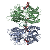
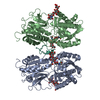
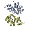
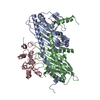
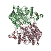
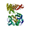
| Title | ROTATION MECHANISM FOR TRANSMEMBRANE SIGNALING BY THE ATRIAL NATRIURETIC PEPTIDE RECEPTOR | |||||||||
 Components Components |
| |||||||||
 Keywords Keywords | SIGNALING PROTEIN / MEMBRANE PROTEIN / RECEPTOR-HORMONE COMPLEX / NATRIURETIC PEPTIDE RECEPTOR / GUANYLYL-CYCLASE-COUPLED RECEPTOR / SIGNAL TRANSDUCTION / ROTATION MECHANISM | |||||||||
| Function / homology |  Function and homology information Function and homology informationPhysiological factors / negative regulation of collecting lymphatic vessel constriction / ANPR-A receptor complex / natriuretic peptide receptor activity / cGMP metabolic process / : / neuropeptide receptor binding / mast cell granule / response to 3-methylcholanthrene / cell growth involved in cardiac muscle cell development ...Physiological factors / negative regulation of collecting lymphatic vessel constriction / ANPR-A receptor complex / natriuretic peptide receptor activity / cGMP metabolic process / : / neuropeptide receptor binding / mast cell granule / response to 3-methylcholanthrene / cell growth involved in cardiac muscle cell development / peptide receptor activity / regulation of body fluid levels / positive regulation of potassium ion export across plasma membrane / guanylate cyclase / cGMP biosynthetic process / receptor guanylyl cyclase signaling pathway / guanylate cyclase activity / negative regulation of JUN kinase activity / synaptic signaling via neuropeptide / regulation of atrial cardiac muscle cell membrane repolarization / multicellular organismal-level water homeostasis / cardiac muscle hypertrophy in response to stress / sodium ion export across plasma membrane / : / neuropeptide hormone activity / hormone receptor binding / regulation of vascular permeability / glycinergic synapse / negative regulation of systemic arterial blood pressure / regulation of calcium ion transmembrane transport via high voltage-gated calcium channel / dopamine metabolic process / hormone binding / cellular response to angiotensin / peptide hormone binding / brush border / negative regulation of epidermal growth factor receptor signaling pathway / positive regulation of heart rate / negative regulation of blood pressure / cell projection / positive regulation of cardiac muscle contraction / blood vessel diameter maintenance / cellular response to glucose stimulus / negative regulation of smooth muscle cell proliferation / female pregnancy / negative regulation of canonical Wnt signaling pathway / cellular response to mechanical stimulus / neuropeptide signaling pathway / hormone activity / response to insulin / negative regulation of cell growth / regulation of blood pressure / vasodilation / cellular response to hydrogen peroxide / heart development / protein folding / perikaryon / response to hypoxia / protein kinase activity / cell surface receptor signaling pathway / signaling receptor complex / intracellular signal transduction / signaling receptor binding / GTP binding / perinuclear region of cytoplasm / protein-containing complex / : / ATP binding / plasma membrane / cytoplasm Similarity search - Function | |||||||||
| Biological species |  | |||||||||
| Method |  X-RAY DIFFRACTION / SYNCHROTRON / MOLECULAR REPLACEMENT / Resolution: 2.95 Å X-RAY DIFFRACTION / SYNCHROTRON / MOLECULAR REPLACEMENT / Resolution: 2.95 Å | |||||||||
 Authors Authors | Ogawa, H. / Qiu, Y. / Ogata, C.M. / Misono, K.S. | |||||||||
 Citation Citation | Journal: J.Biol.Chem. / Year: 2004 Title: Crystal structure of hormone-bound atrial natriuretic peptide receptor extracellular domain: rotation mechanism for transmembrane signal transduction Authors: Ogawa, H. / Qiu, Y. / Ogata, C.M. / Misono, K.S. #1: Journal: Acta Crystallogr.,Sect.D / Year: 2003Title: Crystallization and preliminary x-ray analysis of atrial natriuretic peptide (anp) receptor extracellular domain complex with anp: use of ammonium sulfate as the cryosalt Authors: Ogawa, H. / Zhang, X. / Qiu, Y. / Ogata, C.M. / Misono, K.S. #2: Journal: J.Biol.Chem. / Year: 2004Title: Constitutive activation and uncoupling of the atrial natriuretic peptide receptor by mutations at the dimer interface: role of dimer interface in signaling Authors: Qiu, Y. / Ogawa, H. / Miyagi, M. / Misono, K.S. | |||||||||
| History |
|
- Structure visualization
| Structure viewer | Molecule: MolmilJmol/JSmol |
|---|
- Downloads & links
-Download
| PDBx/mmCIF format | 1t34.cif.gz | 182.2 KB | Display | PDBx/mmCIF format |
|---|---|---|---|---|
| PDB format | pdb1t34.ent.gz | 146.9 KB | Display | PDB format |
| PDBx/mmJSON format | 1t34.json.gz | Tree view | PDBx/mmJSON format | |
| Others |  Other downloads Other downloads |
-Validation report
| Arichive directory | https://data.pdbj.org/pub/pdb/validation_reports/t3/1t34ftp://data.pdbj.org/pub/pdb/validation_reports/t3/1t34 | HTTPS FTP |
|---|
-Related structure data
| Related structure data | 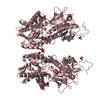 1dp4S S: Starting model for refinement |
|---|---|
| Similar structure data |


-Links
 PDBj
PDBj


- Assembly
| Deposited unit | 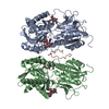
| ||||||||||
|---|---|---|---|---|---|---|---|---|---|---|---|
| 1 |
| ||||||||||
| Unit cell |
|
-Components
| #1: Protein | Mass: 48434.828 Da / Num. of mol.: 2 / Fragment: HORMONE BINDING DOMAIN (RESIDUES 1-435) Source method: isolated from a genetically manipulated source Source: (gene. exp.)  Cricetulus griseus (Chinese hamster) / References: UniProt: P18910 Cricetulus griseus (Chinese hamster) / References: UniProt: P18910#2: Protein/peptide | | Mass: 2217.515 Da / Num. of mol.: 1 / Fragment: RESIDUES 129-149 / Source method: obtained synthetically / Details: This sequence occurs in Rattus norvegicus / References: UniProt: P01161 #3: Polysaccharide | 2-acetamido-2-deoxy-beta-D-glucopyranose-(1-4)-2-acetamido-2-deoxy-beta-D-glucopyranose Source method: isolated from a genetically manipulated source #4: Chemical |   Mass: 35.453 Da / Num. of mol.: 2 / Source method: obtained synthetically / Formula: Cl Mass: 35.453 Da / Num. of mol.: 2 / Source method: obtained synthetically / Formula: ClHas protein modification | Y | |
|---|
-Experimental details
-Experiment
| Experiment | Method: X-RAY DIFFRACTION / Number of used crystals: 1 |
|---|
- Sample preparation
| Crystal | Density Matthews: 3.21 Å3/Da / Density % sol: 67 % |
|---|---|
| Crystal grow | Method: vapor diffusion, hanging drop / pH: 6.5 Details: AMMONIUM SULFATE, SODIUM CHLORIDE, pH 6.5, VAPOR DIFFUSION, HANGING DROP |
-Data collection
| Diffraction | Mean temperature: 100 K |
|---|---|
| Diffraction source | Source: SYNCHROTRON / Site: APS  / Beamline: 19-ID / Wavelength: 0.979338 Å / Beamline: 19-ID / Wavelength: 0.979338 Å |
| Detector | Type: SBC-2 / Detector: CCD / Date: Mar 22, 2003 |
| Radiation | Protocol: SINGLE WAVELENGTH / Monochromatic (M) / Laue (L): M / Scattering type: x-ray |
| Radiation wavelength | Wavelength: 0.979338 Å / Relative weight: 1 |
| Reflection | Resolution: 2.95→87.71 Å / Num. all: 30886 / Num. obs: 25503 / % possible obs: 93.7 % / Observed criterion σ(I): 0.5 / Rmerge(I) obs: 0.054 / Net I/σ(I): 30.9 |
| Reflection shell | Resolution: 2.95→3.04 Å / Rmerge(I) obs: 0.554 / Mean I/σ(I) obs: 3.2 / % possible all: 89.8 |
- Processing
| Software |
| |||||||||||||||||||||||||||||||||||||||||||||||||||||||||||||||||
|---|---|---|---|---|---|---|---|---|---|---|---|---|---|---|---|---|---|---|---|---|---|---|---|---|---|---|---|---|---|---|---|---|---|---|---|---|---|---|---|---|---|---|---|---|---|---|---|---|---|---|---|---|---|---|---|---|---|---|---|---|---|---|---|---|---|---|
| Refinement | Method to determine structure: MOLECULAR REPLACEMENT Starting model: PDB ENTRY 1DP4 Resolution: 2.95→87.71 Å / Cor.coef. Fo:Fc: 0.914 / Cor.coef. Fo:Fc free: 0.902 / SU B: 20.514 / SU ML: 0.378 / Cross valid method: THROUGHOUT / σ(F): 1 / ESU R Free: 0.445 / Stereochemistry target values: MAXIMUM LIKELIHOOD
| |||||||||||||||||||||||||||||||||||||||||||||||||||||||||||||||||
| Solvent computation | Ion probe radii: 0.8 Å / Shrinkage radii: 0.8 Å / VDW probe radii: 1.4 Å / Solvent model: BABINET MODEL WITH MASK | |||||||||||||||||||||||||||||||||||||||||||||||||||||||||||||||||
| Displacement parameters | Biso mean: 52.512 Å2
| |||||||||||||||||||||||||||||||||||||||||||||||||||||||||||||||||
| Refinement step | Cycle: LAST / Resolution: 2.95→87.71 Å
| |||||||||||||||||||||||||||||||||||||||||||||||||||||||||||||||||
| Refine LS restraints |
| |||||||||||||||||||||||||||||||||||||||||||||||||||||||||||||||||
| LS refinement shell | Resolution: 2.95→3.05 Å / Total num. of bins used: 15 /
|
