 Movie
Movie Controller

[English] 日本語
 Yorodumi
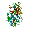
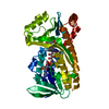
- PDB-6f02: Crystal structure of human glycosylated kallistatin at 3.0 Angstr... -
+ Open data

- Basic information
| Entry | Database: PDB / ID: 6f02 | |||||||||
|---|---|---|---|---|---|---|---|---|---|---|
| Title | Crystal structure of human glycosylated kallistatin at 3.0 Angstrom resolution | |||||||||
 Components Components | Kallistatin | |||||||||
 Keywords Keywords | HYDROLASE / Serine protease inhibitor / kallilrein / protease / inhibitor / heparin | |||||||||
| Function / homology |  Function and homology information Function and homology informationplatelet dense granule lumen / Developmental Lineage of Pancreatic Acinar Cells / serine-type endopeptidase inhibitor activity / Platelet degranulation / : / extracellular exosome / extracellular region Similarity search - Function | |||||||||
| Biological species |  Homo sapiens (human) Homo sapiens (human) | |||||||||
| Method |  X-RAY DIFFRACTION / SYNCHROTRON / MOLECULAR REPLACEMENT / Resolution: 3 Å X-RAY DIFFRACTION / SYNCHROTRON / MOLECULAR REPLACEMENT / Resolution: 3 Å | |||||||||
 Authors Authors | Zhou, A. / Ma, L. | |||||||||
| Funding support |  United Kingdom, United Kingdom,  China, 2items China, 2items
| |||||||||
 Citation Citation | ||||||||||
| History |
|
- Structure visualization
| Structure viewer | Molecule: MolmilJmol/JSmol |
|---|
- Downloads & links
-Download
| PDBx/mmCIF format | 6f02.cif.gz | 314.8 KB | Display | PDBx/mmCIF format |
|---|---|---|---|---|
| PDB format | pdb6f02.ent.gz | 262.2 KB | Display | PDB format |
| PDBx/mmJSON format | 6f02.json.gz | Tree view | PDBx/mmJSON format | |
| Others |  Other downloads Other downloads |
-Validation report
| Arichive directory | https://data.pdbj.org/pub/pdb/validation_reports/f0/6f02ftp://data.pdbj.org/pub/pdb/validation_reports/f0/6f02 | HTTPS FTP |
|---|
-Related structure data
| Related structure data |  6f4uC  6f4vC  1qlpS S: Starting model for refinement C: citing same article ( |
|---|---|
| Similar structure data |


-Links
 PDBj
PDBj
- Assembly
| Deposited unit | 
| ||||||||
|---|---|---|---|---|---|---|---|---|---|
| 1 | 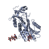
| ||||||||
| 2 | 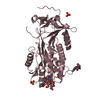
| ||||||||
| Unit cell |
|
-Components
| #1: Protein | Mass: 44595.262 Da / Num. of mol.: 2 Source method: isolated from a genetically manipulated source Details: human Kallistatin was expressed in HEK293 cells with a His6-Tag at the C-terminus Source: (gene. exp.) Homo sapiens (human) / Gene: SERPINA4, KST, PI4 / Cell line (production host): HEK293 / Production host: Homo sapiens (human) / References: UniProt: P29622#2: Polysaccharide | Source method: isolated from a genetically manipulated source #3: Polysaccharide | beta-L-fucopyranose-(1-6)-2-acetamido-2-deoxy-beta-D-glucopyranose | Source method: isolated from a genetically manipulated source #4: Chemical |   Mass: 96.063 Da / Num. of mol.: 2 Mass: 96.063 Da / Num. of mol.: 2Source method: isolated from a genetically manipulated source Formula: SO4 #5: Water | ChemComp-HOH / |  Mass: 18.015 Da / Num. of mol.: 2 / Source method: isolated from a natural source / Formula: H2O Mass: 18.015 Da / Num. of mol.: 2 / Source method: isolated from a natural source / Formula: H2OHas protein modification | Y | |
|---|
-Experimental details
-Experiment
| Experiment | Method: X-RAY DIFFRACTION / Number of used crystals: 1 |
|---|
- Sample preparation
| Crystal | Density Matthews: 4.22 Å3/Da / Density % sol: 70.83 % |
|---|---|
| Crystal grow | Temperature: 293 K / Method: microbatch / Details: PEG3350, Salt |
-Data collection
| Diffraction | Mean temperature: 100 K |
|---|---|
| Diffraction source | Source: SYNCHROTRON / Site: SPring-8  / Beamline: BL12B2 / Wavelength: 0.9785 Å / Beamline: BL12B2 / Wavelength: 0.9785 Å |
| Detector | Type: DECTRIS PILATUS 300K / Detector: PIXEL / Date: Nov 6, 2017 |
| Radiation | Protocol: SINGLE WAVELENGTH / Monochromatic (M) / Laue (L): M / Scattering type: x-ray |
| Radiation wavelength | Wavelength: 0.9785 Å / Relative weight: 1 |
| Reflection | Resolution: 3→104.15 Å / Num. obs: 29350 / % possible obs: 94.6 % / Redundancy: 5.7 % / Net I/σ(I): 9.4 |
| Reflection shell | Resolution: 3→3.18 Å / Redundancy: 5.6 % / Rmerge(I) obs: 0.963 / Mean I/σ(I) obs: 1.3 / Num. unique obs: 4755 / CC1/2: 0.717 / Rpim(I) all: 0.548 / % possible all: 96.7 |
- Processing
| Software |
| ||||||||||||||||||||||||||||||||||||||||||||||||||||||||||||||||||||||||||||||||||||||||||||||||||||||||||||||||||||||||||||||||||||||||||||||||||||||||||||||||||||||||||||||||||||||
|---|---|---|---|---|---|---|---|---|---|---|---|---|---|---|---|---|---|---|---|---|---|---|---|---|---|---|---|---|---|---|---|---|---|---|---|---|---|---|---|---|---|---|---|---|---|---|---|---|---|---|---|---|---|---|---|---|---|---|---|---|---|---|---|---|---|---|---|---|---|---|---|---|---|---|---|---|---|---|---|---|---|---|---|---|---|---|---|---|---|---|---|---|---|---|---|---|---|---|---|---|---|---|---|---|---|---|---|---|---|---|---|---|---|---|---|---|---|---|---|---|---|---|---|---|---|---|---|---|---|---|---|---|---|---|---|---|---|---|---|---|---|---|---|---|---|---|---|---|---|---|---|---|---|---|---|---|---|---|---|---|---|---|---|---|---|---|---|---|---|---|---|---|---|---|---|---|---|---|---|---|---|---|---|
| Refinement | Method to determine structure: MOLECULAR REPLACEMENT Starting model: 1QLP Resolution: 3→104.15 Å / Cor.coef. Fo:Fc: 0.918 / Cor.coef. Fo:Fc free: 0.871 / SU B: 51.794 / SU ML: 0.385 / Cross valid method: THROUGHOUT / ESU R: 1.051 / ESU R Free: 0.406 / Stereochemistry target values: MAXIMUM LIKELIHOOD / Details: HYDROGENS HAVE BEEN ADDED IN THE RIDING POSITIONS
| ||||||||||||||||||||||||||||||||||||||||||||||||||||||||||||||||||||||||||||||||||||||||||||||||||||||||||||||||||||||||||||||||||||||||||||||||||||||||||||||||||||||||||||||||||||||
| Solvent computation | Ion probe radii: 0.8 Å / Shrinkage radii: 0.8 Å / VDW probe radii: 1.2 Å / Solvent model: MASK | ||||||||||||||||||||||||||||||||||||||||||||||||||||||||||||||||||||||||||||||||||||||||||||||||||||||||||||||||||||||||||||||||||||||||||||||||||||||||||||||||||||||||||||||||||||||
| Displacement parameters | Biso mean: 86.632 Å2
| ||||||||||||||||||||||||||||||||||||||||||||||||||||||||||||||||||||||||||||||||||||||||||||||||||||||||||||||||||||||||||||||||||||||||||||||||||||||||||||||||||||||||||||||||||||||
| Refinement step | Cycle: 1 / Resolution: 3→104.15 Å
| ||||||||||||||||||||||||||||||||||||||||||||||||||||||||||||||||||||||||||||||||||||||||||||||||||||||||||||||||||||||||||||||||||||||||||||||||||||||||||||||||||||||||||||||||||||||
| Refine LS restraints |
|