 Movie
Movie Controller

[English] 日本語
 Yorodumi
- PDB-6f4u: Crystal structure of reactive loop cleaved kallistatin at 1.9 ang... -
+ Open data

- Basic information
| Entry | Database: PDB / ID: 6f4u | |||||||||
|---|---|---|---|---|---|---|---|---|---|---|
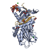
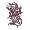
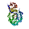
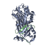
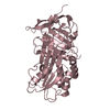
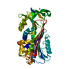
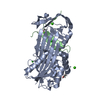
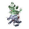
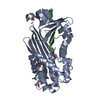
| Title | Crystal structure of reactive loop cleaved kallistatin at 1.9 angstrom resolution | |||||||||
 Components Components | (Kallistatin) x 2 | |||||||||
 Keywords Keywords | HYDROLASE / inhibitor / Serpin / kallekrein / kallistatin | |||||||||
| Function / homology |  Function and homology information Function and homology informationplatelet dense granule lumen / Developmental Lineage of Pancreatic Acinar Cells / serine-type endopeptidase inhibitor activity / Platelet degranulation / : / extracellular exosome / extracellular region Similarity search - Function | |||||||||
| Biological species |  Homo sapiens (human) Homo sapiens (human) | |||||||||
| Method |  X-RAY DIFFRACTION / SYNCHROTRON / Resolution: 1.9 Å X-RAY DIFFRACTION / SYNCHROTRON / Resolution: 1.9 Å | |||||||||
 Authors Authors | Zhou, A. / Wei, Z. / Lin, F. | |||||||||
| Funding support |  United Kingdom, United Kingdom,  China, 2items China, 2items
| |||||||||
 Citation Citation | Journal: To Be Published Title: Crystal structure of human Kallistatin Authors: Zhou, A. / Ma, L. | |||||||||
| History |
|
- Structure visualization
| Structure viewer | Molecule: MolmilJmol/JSmol |
|---|
- Downloads & links
-Download
| PDBx/mmCIF format | 6f4u.cif.gz | 169.9 KB | Display | PDBx/mmCIF format |
|---|---|---|---|---|
| PDB format | pdb6f4u.ent.gz | 133.6 KB | Display | PDB format |
| PDBx/mmJSON format | 6f4u.json.gz | Tree view | PDBx/mmJSON format | |
| Others |  Other downloads Other downloads |
-Validation report
| Arichive directory | https://data.pdbj.org/pub/pdb/validation_reports/f4/6f4uftp://data.pdbj.org/pub/pdb/validation_reports/f4/6f4u | HTTPS FTP |
|---|
-Related structure data


-Links
 PDBj
PDBj
- Assembly
| Deposited unit | 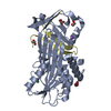
| ||||||||
|---|---|---|---|---|---|---|---|---|---|
| 1 |
| ||||||||
| Unit cell |
|
-Components
-Protein / Protein/peptide , 2 types, 2 molecules AD
| #1: Protein | Mass: 39031.922 Da / Num. of mol.: 1 Source method: isolated from a genetically manipulated source Details: reactive loop cleaved human kallistatin with an Extra Gly at the N-terminus Source: (gene. exp.) Homo sapiens (human) / Gene: SERPINA4, KST, PI4 / Plasmid: Psumo3-Kal / Production host:  |
|---|---|
| #2: Protein/peptide | Mass: 4568.324 Da / Num. of mol.: 1 Source method: isolated from a genetically manipulated source Details: reactive loop cleaved human kallistatin with an Extra Gly at the N-terminus Source: (gene. exp.) Homo sapiens (human) / Gene: SERPINA4, KST, PI4 / Plasmid: Psumo3-Kal / Production host: |
-Non-polymers , 5 types, 201 molecules 


| #3: Chemical | ChemComp-EDO /  Mass: 62.068 Da / Num. of mol.: 7 / Source method: obtained synthetically / Formula: C2H6O2 Mass: 62.068 Da / Num. of mol.: 7 / Source method: obtained synthetically / Formula: C2H6O2#4: Chemical |  Mass: 92.094 Da / Num. of mol.: 2 / Source method: obtained synthetically / Formula: C3H8O3 Mass: 92.094 Da / Num. of mol.: 2 / Source method: obtained synthetically / Formula: C3H8O3#5: Chemical | ChemComp-CL / |  Mass: 35.453 Da / Num. of mol.: 1 / Source method: obtained synthetically / Formula: Cl Mass: 35.453 Da / Num. of mol.: 1 / Source method: obtained synthetically / Formula: Cl#6: Chemical | ChemComp-NA / |  Mass: 22.990 Da / Num. of mol.: 1 / Source method: obtained synthetically / Formula: Na Mass: 22.990 Da / Num. of mol.: 1 / Source method: obtained synthetically / Formula: Na#7: Water | ChemComp-HOH / | Mass: 18.015 Da / Num. of mol.: 190 / Source method: isolated from a natural source / Formula: H2O |
|---|
-Experimental details
-Experiment
| Experiment | Method: X-RAY DIFFRACTION / Number of used crystals: 1 |
|---|
- Sample preparation
| Crystal | Density Matthews: 3.25 Å3/Da / Density % sol: 63 % |
|---|---|
| Crystal grow | Temperature: 293 K / Method: vapor diffusion, sitting drop / Details: 30% tertbutanal |
-Data collection
| Diffraction | Mean temperature: 100 K |
|---|---|
| Diffraction source | Source: SYNCHROTRON / Site: SSRF / Beamline: BL19U1 / Wavelength: 1.196 Å |
| Detector | Type: DECTRIS PILATUS3 X CdTe 1M / Detector: PIXEL / Date: Jan 1, 2011 |
| Radiation | Protocol: SINGLE WAVELENGTH / Monochromatic (M) / Laue (L): M / Scattering type: x-ray |
| Radiation wavelength | Wavelength: 1.196 Å / Relative weight: 1 |
| Reflection | Resolution: 1.9→35.67 Å / Num. obs: 44414 / % possible obs: 99.9 % / Redundancy: 4.2 % / Biso Wilson estimate: 30.1 Å2 / Rmerge(I) obs: 0.043 / Net I/σ(I): 14 |
| Reflection shell | Resolution: 1.9→2 Å / Redundancy: 4.1 % / Rmerge(I) obs: 0.326 / Mean I/σ(I) obs: 2.5 / Num. unique obs: 6433 / % possible all: 99.7 |
- Processing
| Software |
| ||||||||||||||||||||||||||||||||||||||||||||||||||||||||||||||||||||||||||||||||||||||||||||||||||||||||||||||||||||||||||||||||||||||||||||||||||||||||||||||||||||||||||||||||||||||
|---|---|---|---|---|---|---|---|---|---|---|---|---|---|---|---|---|---|---|---|---|---|---|---|---|---|---|---|---|---|---|---|---|---|---|---|---|---|---|---|---|---|---|---|---|---|---|---|---|---|---|---|---|---|---|---|---|---|---|---|---|---|---|---|---|---|---|---|---|---|---|---|---|---|---|---|---|---|---|---|---|---|---|---|---|---|---|---|---|---|---|---|---|---|---|---|---|---|---|---|---|---|---|---|---|---|---|---|---|---|---|---|---|---|---|---|---|---|---|---|---|---|---|---|---|---|---|---|---|---|---|---|---|---|---|---|---|---|---|---|---|---|---|---|---|---|---|---|---|---|---|---|---|---|---|---|---|---|---|---|---|---|---|---|---|---|---|---|---|---|---|---|---|---|---|---|---|---|---|---|---|---|---|---|
| Refinement | Resolution: 1.9→98.5 Å / Cor.coef. Fo:Fc: 0.966 / Cor.coef. Fo:Fc free: 0.957 / SU B: 5.288 / SU ML: 0.076 / Cross valid method: THROUGHOUT / ESU R: 0.11 / ESU R Free: 0.106 / Stereochemistry target values: MAXIMUM LIKELIHOOD / Details: HYDROGENS HAVE BEEN ADDED IN THE RIDING POSITIONS
| ||||||||||||||||||||||||||||||||||||||||||||||||||||||||||||||||||||||||||||||||||||||||||||||||||||||||||||||||||||||||||||||||||||||||||||||||||||||||||||||||||||||||||||||||||||||
| Solvent computation | Ion probe radii: 0.8 Å / Shrinkage radii: 0.8 Å / VDW probe radii: 1.2 Å / Solvent model: MASK | ||||||||||||||||||||||||||||||||||||||||||||||||||||||||||||||||||||||||||||||||||||||||||||||||||||||||||||||||||||||||||||||||||||||||||||||||||||||||||||||||||||||||||||||||||||||
| Displacement parameters | Biso mean: 34.154 Å2
| ||||||||||||||||||||||||||||||||||||||||||||||||||||||||||||||||||||||||||||||||||||||||||||||||||||||||||||||||||||||||||||||||||||||||||||||||||||||||||||||||||||||||||||||||||||||
| Refinement step | Cycle: 1 / Resolution: 1.9→98.5 Å
| ||||||||||||||||||||||||||||||||||||||||||||||||||||||||||||||||||||||||||||||||||||||||||||||||||||||||||||||||||||||||||||||||||||||||||||||||||||||||||||||||||||||||||||||||||||||
| Refine LS restraints |
|