[English] 日本語
Yorodumi
- PDB-7bp3: Cryo-EM structure of the human MCT2 -

+
Open data


ID or keywords:

Loading...

-
Basic information

Entry
Database: PDB / ID: 7bp3
TitleCryo-EM structure of the human MCT2
ComponentsMonocarboxylate transporter 2
KeywordsTRANSPORT PROTEIN / Monocarboxylate transporter 2 / Major facilitator superfamily / Cooperative transport
Function / homology
Function and homology information


pyruvate secondary active transmembrane transporter activity / pyruvate transmembrane transporter activity / lactate transmembrane transport / pyruvate transmembrane transport / monocarboxylic acid transmembrane transporter activity / lactate transmembrane transporter activity / monocarboxylic acid transport / Proton-coupled monocarboxylate transport / symporter activity / plasma membrane => GO:0005886 ...pyruvate secondary active transmembrane transporter activity / pyruvate transmembrane transporter activity / lactate transmembrane transport / pyruvate transmembrane transport / monocarboxylic acid transmembrane transporter activity / lactate transmembrane transporter activity / monocarboxylic acid transport / Proton-coupled monocarboxylate transport / symporter activity / plasma membrane => GO:0005886 / transport across blood-brain barrier / membrane => GO:0016020 / nucleoplasm / plasma membrane
Similarity search - Function
Monocarboxylate transporter 2 / Monocarboxylate transporter / Major facilitator superfamily / Major Facilitator Superfamily / Major facilitator superfamily domain / Major facilitator superfamily (MFS) profile. / MFS transporter superfamily
Similarity search - Domain/homology
Monocarboxylate transporter 2
Similarity search - Component
Biological speciesHomo sapiens (human)
MethodELECTRON MICROSCOPY / single particle reconstruction / cryo EM / Resolution: 3.8 Å
AuthorsZhang, B. / Jin, Q. / Zhang, X. / Guo, J. / Ye, S.
Citation
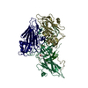
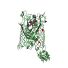
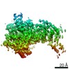
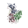
Journal: Nat Commun / Year: 2020
Title: Cooperative transport mechanism of human monocarboxylate transporter 2.
Authors: Bo Zhang / Qiuheng Jin / Lizhen Xu / Ningning Li / Ying Meng / Shenghai Chang / Xiang Zheng / Jiangqin Wang / Yuan Chen / Dante Neculai / Ning Gao / Xiaokang Zhang / Fan Yang / Jiangtao Guo / Sheng Ye /
Abstract: Proton-linked monocarboxylate transporters (MCTs) must transport monocarboxylate efficiently to facilitate monocarboxylate efflux in glycolytically active cells, and transport monocarboxylate slowly ...Proton-linked monocarboxylate transporters (MCTs) must transport monocarboxylate efficiently to facilitate monocarboxylate efflux in glycolytically active cells, and transport monocarboxylate slowly or even shut down to maintain a physiological monocarboxylate concentration in glycolytically inactive cells. To discover how MCTs solve this fundamental aspect of intracellular monocarboxylate homeostasis in the context of multicellular organisms, we analyzed pyruvate transport activity of human monocarboxylate transporter 2 (MCT2). Here we show that MCT2 transport activity exhibits steep dependence on substrate concentration. This property allows MCTs to turn on almost like a switch, which is physiologically crucial to the operation of MCTs in the cellular context. We further determined the cryo-electron microscopy structure of the human MCT2, demonstrating that the concentration sensitivity of MCT2 arises from the strong inter-subunit cooperativity of the MCT2 dimer during transport. These data establish definitively a clear example of evolutionary optimization of protein function.
History
DepositionMar 21, 2020Deposition site: PDBJ / Processing site: PDBJ
Revision 1.0Jun 3, 2020Provider: repository / Type: Initial release
Revision 1.1Mar 27, 2024Group: Data collection / Database references / Category: chem_comp_atom / chem_comp_bond / database_2
Item: _database_2.pdbx_DOI / _database_2.pdbx_database_accession

-
Structure visualization

Movie
  • Deposited structure unit
  • Imaged by Jmol
  • Download
  • Simplified surface model + fitted atomic model
  • EMDB-30143
  • Imaged by Jmol
  • Download
  • Superimposition on EM map
  • EMDB-30143
  • Imaged by UCSF Chimera
  • Download
Movie viewer
Structure viewerMolecule:
MolmilJmol/JSmol

Downloads & links

-
Assembly

Deposited unit
A: Monocarboxylate transporter 2
B: Monocarboxylate transporter 2


Theoretical massNumber of molelcules
Total (without water)107,8392
Polymers107,8392
Non-polymers00
Water0
1


  • Idetical with deposited unit
  • defined by author
  • Evidence: gel filtration
TypeNameSymmetry operationNumber
identity operation1_5551
Buried area5080 Å2
ΔGint-54 kcal/mol
Surface area33690 Å2

-
Components

#1: Protein Monocarboxylate transporter 2 / / MCT 2 / Solute carrier family 16 member 7


Mass: 53919.707 Da / Num. of mol.: 2
Source method: isolated from a genetically manipulated source
Source: (gene. exp.) Homo sapiens (human) / Gene: SLC16A7, MCT2 / Cell line (production host): HEK293F / Production host: Homo sapiens (human) / References: UniProt: O60669

-
Experimental details

-
Experiment

ExperimentMethod: ELECTRON MICROSCOPY
EM experimentAggregation state: PARTICLE / 3D reconstruction method: single particle reconstruction

-
Sample preparation

ComponentName: Monocarboxylate transporter 2 / Type: COMPLEX / Entity ID: all / Source: RECOMBINANT
Source (natural)Organism: Homo sapiens (human)
Source (recombinant)Organism: Homo sapiens (human)
Buffer solutionpH: 7.5
Buffer component
IDConc.NameFormulaBuffer-ID
150 mM4-(2-hydroxyethyl)-1-piperazineethanesulfonic acid4-(2-Hydroxyethyl)-1-piperazineethanesulfonic acidHEPES1
2150 mMsodium chlorideNaClSodium chloride1
SpecimenConc.: 4 mg/ml / Embedding applied: NO / Shadowing applied: NO / Staining applied: NO / Vitrification applied: YES
Specimen supportGrid material: GOLD / Grid mesh size: 300 divisions/in. / Grid type: Quantifoil R1.2/1.3
VitrificationInstrument: FEI VITROBOT MARK IV / Cryogen name: ETHANE / Humidity: 100 % / Chamber temperature: 277 K / Details: blot for 4 seconds before plunging

-
Electron microscopy imaging

Experimental equipment
Model: Titan Krios / Image courtesy: FEI Company
MicroscopyModel: FEI TITAN KRIOS
Electron gunElectron source: FIELD EMISSION GUN / Accelerating voltage: 300 kV / Illumination mode: FLOOD BEAM
Electron lensMode: BRIGHT FIELDBright-field microscopy
Image recordingAverage exposure time: 0.25 sec. / Electron dose: 82 e/Å2 / Film or detector model: GATAN K2 SUMMIT (4k x 4k)
Image scansMovie frames/image: 32

-
Processing

EM software
IDNameVersionCategory
1RELION3particle selection
4RELION3CTF correction
7Coot0.89model fitting
9PHENIX1.14model refinement
13cryoSPARC23D reconstruction
CTF correctionType: PHASE FLIPPING AND AMPLITUDE CORRECTION
Particle selectionNum. of particles selected: 302904
SymmetryPoint symmetry: C2 (2 fold cyclic)
3D reconstructionResolution: 3.8 Å / Resolution method: FSC 0.143 CUT-OFF / Num. of particles: 100909 / Num. of class averages: 1 / Symmetry type: POINT
Atomic model buildingProtocol: AB INITIO MODEL / Space: REAL

+
About Yorodumi

-
News

-
Feb 9, 2022. New format data for meta-information of EMDB entries

New format data for meta-information of EMDB entries

  • Version 3 of the EMDB header file is now the official format.
  • The previous official version 1.9 will be removed from the archive.

Related info.:EMDB header

External links:wwPDB to switch to version 3 of the EMDB data model

-
Aug 12, 2020. Covid-19 info

Covid-19 info

URL: https://pdbj.org/emnavi/covid19.php

New page: Covid-19 featured information page in EM Navigator.

Related info.:Covid-19 info / Mar 5, 2020. Novel coronavirus structure data

+
Mar 5, 2020. Novel coronavirus structure data

Novel coronavirus structure data

Related info.:Yorodumi Speices / Aug 12, 2020. Covid-19 info

External links:COVID-19 featured content - PDBj / Molecule of the Month (242):Coronavirus Proteases

+
Jan 31, 2019. EMDB accession codes are about to change! (news from PDBe EMDB page)

EMDB accession codes are about to change! (news from PDBe EMDB page)

  • The allocation of 4 digits for EMDB accession codes will soon come to an end. Whilst these codes will remain in use, new EMDB accession codes will include an additional digit and will expand incrementally as the available range of codes is exhausted. The current 4-digit format prefixed with “EMD-” (i.e. EMD-XXXX) will advance to a 5-digit format (i.e. EMD-XXXXX), and so on. It is currently estimated that the 4-digit codes will be depleted around Spring 2019, at which point the 5-digit format will come into force.
  • The EM Navigator/Yorodumi systems omit the EMD- prefix.

Related info.:Q: What is EMD? / ID/Accession-code notation in Yorodumi/EM Navigator

External links:EMDB Accession Codes are Changing Soon! / Contact to PDBj

+
Jul 12, 2017. Major update of PDB

Major update of PDB

  • wwPDB released updated PDB data conforming to the new PDBx/mmCIF dictionary.
  • This is a major update changing the version number from 4 to 5, and with Remediation, in which all the entries are updated.
  • In this update, many items about electron microscopy experimental information are reorganized (e.g. em_software).
  • Now, EM Navigator and Yorodumi are based on the updated data.

External links:wwPDB Remediation / Enriched Model Files Conforming to OneDep Data Standards Now Available in the PDB FTP Archive

-
Yorodumi

Thousand views of thousand structures

  • Yorodumi is a browser for structure data from EMDB, PDB, SASBDB, etc.
  • This page is also the successor to EM Navigator detail page, and also detail information page/front-end page for Omokage search.
  • The word "yorodu" (or yorozu) is an old Japanese word meaning "ten thousand". "mi" (miru) is to see.

Related info.:EMDB / PDB / SASBDB / Comparison of 3 databanks / Yorodumi Search / Aug 31, 2016. New EM Navigator & Yorodumi / Yorodumi Papers / Jmol/JSmol / Function and homology information / Changes in new EM Navigator and Yorodumi

Read more