 Movie
Movie Controller

[English] 日本語
 Yorodumi
- EMDB-4724: Structure of a membrane adenylyl cyclase bound to an activated st... -
+ Open data

- Basic information
| Entry | Database: EMDB / ID: EMD-4724 | |||||||||
|---|---|---|---|---|---|---|---|---|---|---|
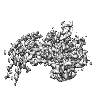
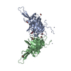
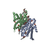
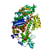
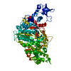
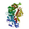
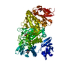
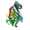
| Title | Structure of a membrane adenylyl cyclase bound to an activated stimulatory G protein (SOL-C map) | |||||||||
 Map data Map data | Soluble domain of AC9 bound to GalphaS (map SOL-C) | |||||||||
 Sample Sample |
| |||||||||
| Function / homology |  Function and homology information Function and homology informationAdenylate cyclase activating pathway / Adenylate cyclase inhibitory pathway / PKA activation / adenylate cyclase / Hedgehog 'off' state / sensory perception of chemical stimulus / mu-type opioid receptor binding / corticotropin-releasing hormone receptor 1 binding / adenylate cyclase activity / cAMP biosynthetic process ...Adenylate cyclase activating pathway / Adenylate cyclase inhibitory pathway / PKA activation / adenylate cyclase / Hedgehog 'off' state / sensory perception of chemical stimulus / mu-type opioid receptor binding / corticotropin-releasing hormone receptor 1 binding / adenylate cyclase activity / cAMP biosynthetic process / beta-2 adrenergic receptor binding / G alpha (z) signalling events / D1 dopamine receptor binding / adenylate cyclase-activating adrenergic receptor signaling pathway / insulin-like growth factor receptor binding / ionotropic glutamate receptor binding / adenylate cyclase activator activity / G-protein beta/gamma-subunit complex binding / adenylate cyclase-activating dopamine receptor signaling pathway / heterotrimeric G-protein complex / adenylate cyclase-activating G protein-coupled receptor signaling pathway / in utero embryonic development / Hydrolases; Acting on acid anhydrides; Acting on GTP to facilitate cellular and subcellular movement / intracellular signal transduction / ciliary basal body / GTPase activity / GTP binding / ATP binding / metal ion binding / plasma membrane / cytoplasm / cytosol Similarity search - Function | |||||||||
| Biological species |  | |||||||||
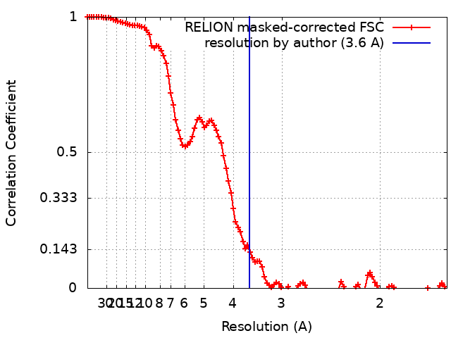
| Method | single particle reconstruction / cryo EM / Resolution: 3.6 Å | |||||||||
 Authors Authors | Korkhov VM / Qi C | |||||||||
| Funding support |  Switzerland, 1 items Switzerland, 1 items
| |||||||||
 Citation Citation | Journal: Science / Year: 2019 Title: The structure of a membrane adenylyl cyclase bound to an activated stimulatory G protein. Authors: Chao Qi / Simona Sorrentino / Ohad Medalia / Volodymyr M Korkhov /  Abstract: Membrane-integral adenylyl cyclases (ACs) are key enzymes in mammalian heterotrimeric GTP-binding protein (G protein)-dependent signal transduction, which is important in many cellular processes. ...Membrane-integral adenylyl cyclases (ACs) are key enzymes in mammalian heterotrimeric GTP-binding protein (G protein)-dependent signal transduction, which is important in many cellular processes. Signals received by the G protein-coupled receptors are conveyed to ACs through G proteins to modulate the levels of cellular cyclic adenosine monophosphate (cAMP). Here, we describe the cryo-electron microscopy structure of the bovine membrane AC9 bound to an activated G protein αs subunit at 3.4-angstrom resolution. The structure reveals the organization of the membrane domain and helical domain that spans between the membrane and catalytic domains of AC9. The carboxyl-terminal extension of the catalytic domain occludes both the catalytic and the allosteric sites of AC9, inducing a conformation distinct from the substrate- and activator-bound state, suggesting a regulatory role in cAMP production. | |||||||||
| History |
|
- Structure visualization
| Movie |
Movie viewer |
|---|---|
| Structure viewer | EM map: SurfViewMolmilJmol/JSmol |
| Supplemental images |
- Downloads & links
-EMDB archive
| Map data | emd_4724.map.gz | 7.7 MB | EMDB map data format | |
|---|---|---|---|---|
| Header (meta data) | emd-4724-v30.xmlemd-4724.xml | 16.4 KB 16.4 KB | Display Display | EMDB header |
| FSC (resolution estimation) | emd_4724_fsc.xml | 10.7 KB | Display | FSC data file |
| Images |  emd_4724.png emd_4724.png | 52.5 KB | ||
| Archive directory |  http://ftp.pdbj.org/pub/emdb/structures/EMD-4724ftp://ftp.pdbj.org/pub/emdb/structures/EMD-4724 http://ftp.pdbj.org/pub/emdb/structures/EMD-4724ftp://ftp.pdbj.org/pub/emdb/structures/EMD-4724 | HTTPS FTP |
-Related structure data
| Related structure data | 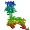 4719C 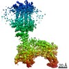 4721C 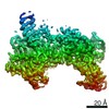 4722C 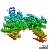 4723C 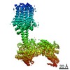 4725C 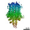 4726C  6r3qC 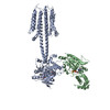 6r4oC 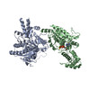 6r4pC C: citing same article ( |
|---|---|
| Similar structure data |

-Links
| EMDB pages | EMDB (EBI/PDBe) / EMDataResource |
|---|---|
| Related items in Molecule of the Month |


-Map
| File | Download / File: emd_4724.map.gz / Format: CCP4 / Size: 103 MB / Type: IMAGE STORED AS FLOATING POINT NUMBER (4 BYTES) | ||||||||||||||||||||||||||||||||||||||||||||||||||||||||||||
|---|---|---|---|---|---|---|---|---|---|---|---|---|---|---|---|---|---|---|---|---|---|---|---|---|---|---|---|---|---|---|---|---|---|---|---|---|---|---|---|---|---|---|---|---|---|---|---|---|---|---|---|---|---|---|---|---|---|---|---|---|---|
| Annotation | Soluble domain of AC9 bound to GalphaS (map SOL-C) | ||||||||||||||||||||||||||||||||||||||||||||||||||||||||||||
| Projections & slices | Image control
Images are generated by Spider. | ||||||||||||||||||||||||||||||||||||||||||||||||||||||||||||
| Voxel size | X=Y=Z: 0.814 Å | ||||||||||||||||||||||||||||||||||||||||||||||||||||||||||||
| Density |
| ||||||||||||||||||||||||||||||||||||||||||||||||||||||||||||
| Symmetry | Space group: 1 | ||||||||||||||||||||||||||||||||||||||||||||||||||||||||||||
| Details | EMDB XML:
CCP4 map header:
| ||||||||||||||||||||||||||||||||||||||||||||||||||||||||||||
 Z (Sec.)
 Y (Row.)
 X (Col.)


-Supplemental data
- Sample components
-Entire : Complex of adenylyl cyclase AC9 with G protein subunit Galphas
| Entire | Name: Complex of adenylyl cyclase AC9 with G protein subunit Galphas |
|---|---|
| Components |
|
-Supramolecule #1: Complex of adenylyl cyclase AC9 with G protein subunit Galphas
| Supramolecule | Name: Complex of adenylyl cyclase AC9 with G protein subunit Galphas type: complex / ID: 1 / Parent: 0 / Macromolecule list: all |
|---|
-Supramolecule #2: Adenylate cyclase 9
| Supramolecule | Name: Adenylate cyclase 9 / type: complex / ID: 2 / Parent: 1 / Macromolecule list: #1 |
|---|---|
| Source (natural) | Organism: |
| Recombinant expression | Organism:  Homo sapiens (human) / Recombinant cell: HEK293F / Recombinant plasmid: pEZT-BM Homo sapiens (human) / Recombinant cell: HEK293F / Recombinant plasmid: pEZT-BM |
-Supramolecule #3: Guanine nucleotide-binding protein G(s) subunit alpha isoforms short
| Supramolecule | Name: Guanine nucleotide-binding protein G(s) subunit alpha isoforms short type: complex / ID: 3 / Parent: 1 / Macromolecule list: #2 |
|---|---|
| Source (natural) | Organism: |
| Recombinant expression | Organism:  Trichoplusia ni (cabbage looper) / Recombinant cell: High Five / Recombinant plasmid: pFastbac Trichoplusia ni (cabbage looper) / Recombinant cell: High Five / Recombinant plasmid: pFastbac |
-Macromolecule #1: Adenylyl cylcase AC9
| Macromolecule | Name: Adenylyl cylcase AC9 / type: protein_or_peptide / ID: 1 / Enantiomer: LEVO / EC number: adenylate cyclase |
|---|---|
| Source (natural) | Organism: |
| Recombinant expression | Organism: Homo sapiens (human) |
| Sequence | String: MASPPHQQLL QHHSTEVSCD SSGDSNSVRV RINPKQPSSN SHPKHCKYSI SSSCSSSGDS GGVPRRMGAG GRLRRRKKLP QLFERASSRW WDPKFDSVNL EEACMERCFP QTQRRFRYAL FYIGFACLLW SIYFGVHMKS KLIVMVAPAL CFLVVCVGFF LFTFTKLYAR ...String: MASPPHQQLL QHHSTEVSCD SSGDSNSVRV RINPKQPSSN SHPKHCKYSI SSSCSSSGDS GGVPRRMGAG GRLRRRKKLP QLFERASSRW WDPKFDSVNL EEACMERCFP QTQRRFRYAL FYIGFACLLW SIYFGVHMKS KLIVMVAPAL CFLVVCVGFF LFTFTKLYAR HYVWTSLVLT LLVFALTLAA QFQVLTPLSG RVDNFNHTRA ARPTDTCLSQ VGSFSMCIEV LFLLYTVMHL PLYLSLILGV AYSVLFETFG YHFQDEACFA SPGAEALHWE LLSRALLHLC IHAIGIHLFI MSQVRSRSTF LKVGQSIMHG KDLEVEKALK ERMIHSVMPR IIADDLMKQG DEESENSVKR HATSSPKNRK KKSSIQKAPI AFRPFKMQQI EEVSILFADI VGFTKMSANK SAHALVGLLN DLFGRFDRLC EETKCEKIST LGDCYYCVAG CPEPRADHAY CCIEMGLGMI RAIEQFCQEK KEMVNMRVGV HTGTVLCGIL GMRRFKFDVW SNDVNLANLM EQLGVAGKVH ISEATAKYLD DRYEMEDGKV TERLGQSVVA DQLKGLKTYL IAGQRAKESH CSCSEALLSG FEVLDGSRVS SGPRGQGTAS PGSVSDLAQT VKTFDNLKTC PSCGITFTPK PEAGAEGGAV QNGCQEEPKN SAKASGGPSS KTQNGLLSPP PEEKLTNSQT SLCEILQEKG RWAGVSLDQS ALLPLRFKNI REKTDAHFVD VIKEDSLMKD YFFKPPINQF SLNFLDPELE RAYRTSYQEE VVKSSPVRTF ASATFSSLLD VLLSTTVFLI LSITCFLRYG AASTPPPPAA LAVFGAALLL EILSLVVSVR MVFFLEDVMT CTKRLLEWIA GWLPRHFIGA ILVSLPALAV YSHVTSEFET NIHSTMFTGS AVLTAVVQYC NFCQLSSWMR SSLATVVGAG PLLLLLYVSL CPDSSTVISH LDAVQNFSST RKLCNASLPH DGRSPASLIG QEVILVFFLL LLLVWFLNRE FEVSYRLHYH GDVEADLHRT KIQSMRDQAD WLLRNIIPYH VAEQLKVSQT YSKNHDSGGV IFASIVNFSE FYEENYEGGK ECYRVLNELI GDFDELLSKP DYSSIEKIKT IGATYMAASG LNATQCRDGS HPQEHLQILF EFAKEMMRVV DDFNNNMLWF NFKLRVGFNH GPLTAGVIGT TKLLYDIWGD TVNIASRMDT TGVECRIQVS EESYRVLSKM GYEFDYRGTV NVKGKGQMKT YLYPKCTDSG LVPQHQLSIS PDIRVQVDGS IGRSPTDEIA SLVPSVQNPD QVPPGSENNA QTRDAHPSAK RPWKEPVRAE ERCRFGKAIE KSDCEEVGME EANELTKLNV SERAAAALEV LFQGPGGVSK GEELFTGVVP ILVELDGDVN GHKFSVSGEG EGDATYGKLT LKFICTTGKL PVPWPTLVTT FGYGLQCFAR YPDHMKQHDF FKSAMPEGYV QERTIFFKDD GNYKTRAEVK FEGDTLVNRI ELKGIDFKED GNILGHKLEY NYNSHNVYIM ADKQKNGIKV NFKIRHNIED GSVQLADHYQ QNTPIGDGPV LLPDNHYLSY QSALSKDPNE KRDHMVLLEF VTAAGITLGM DELYKAASAW SHPQFEKGGG SGGGSGGSAW SHPQFEK |
-Macromolecule #2: Guanine nucleotide-binding protein G(s) subunit alpha isoforms short
| Macromolecule | Name: Guanine nucleotide-binding protein G(s) subunit alpha isoforms short type: protein_or_peptide / ID: 2 / Enantiomer: LEVO |
|---|---|
| Source (natural) | Organism: |
| Recombinant expression | Organism: Trichoplusia ni (cabbage looper) |
| Sequence | String: MGCLGNSKTE DQRNEEKAQR EANKKIEKQL QKDKQVYRAT HRLLLLGAGE SGKSTIVKQM RILHVNGFNG GEGGEEDPNA KSNSDGEKAT KVQDIKNNLK EAIETIVAAM SNLVPPVELA NPENQFRVDY ILSVMNVPDF DFPPEFYEHA KALWEDEGVR ACYERSNEYQ ...String: MGCLGNSKTE DQRNEEKAQR EANKKIEKQL QKDKQVYRAT HRLLLLGAGE SGKSTIVKQM RILHVNGFNG GEGGEEDPNA KSNSDGEKAT KVQDIKNNLK EAIETIVAAM SNLVPPVELA NPENQFRVDY ILSVMNVPDF DFPPEFYEHA KALWEDEGVR ACYERSNEYQ LIDCAQYFLD KIDVIKQDDY VPSDQDLLRC RVLTSGIFET KFQVDKVNFH MFDVGGQRDE RRKWIQCFND VTAIIFVVAS SSYNMVIRED NQTNRLQEAL NLFKSIWNNR WLRTISVILF LNKQDLLAEK VLAGKSKIED YFPEFARYTT PEDATPEPGE DPRVTRAKYF IRDEFLRIST ASGDGRHYCY PHFTCAVDTE NIRRVFNDCR DIIQRMHLRQ YELLGGHHHH HHHH |
-Experimental details
-Structure determination
| Method | cryo EM |
|---|---|
 Processing Processing | single particle reconstruction |
| Aggregation state | particle |
-Sample preparation
| Buffer | pH: 8 |
|---|---|
| Vitrification | Cryogen name: ETHANE / Chamber humidity: 100 % / Chamber temperature: 278 K / Instrument: FEI VITROBOT MARK IV |
- Electron microscopy
| Microscope | FEI TITAN KRIOS |
|---|---|
| Image recording | Film or detector model: GATAN K2 QUANTUM (4k x 4k) / Detector mode: SUPER-RESOLUTION / Number real images: 5817 / Average exposure time: 8.0 sec. / Average electron dose: 47.0 e/Å2 |
| Electron beam | Acceleration voltage: 300 kV / Electron source:  FIELD EMISSION GUN FIELD EMISSION GUN |
| Electron optics | Illumination mode: FLOOD BEAM / Imaging mode: BRIGHT FIELD / Nominal defocus max: 2.5 µm / Nominal defocus min: 0.75 µm |
| Sample stage | Specimen holder model: FEI TITAN KRIOS AUTOGRID HOLDER / Cooling holder cryogen: NITROGEN |
| Experimental equipment |  Model: Titan Krios / Image courtesy: FEI Company |
+Image processing
-Atomic model buiding 1
| Refinement | Space: REAL / Protocol: AB INITIO MODEL / Overall B value: 149.61 / Target criteria: Cross-correlation coefficient |
|---|
