Movie
Movie Controller
 Structure viewers
 About Yorodumi Papers


+Search query
-Structure paper
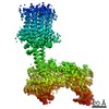
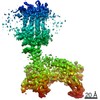
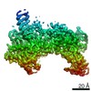
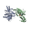
| Title | The structure of a membrane adenylyl cyclase bound to an activated stimulatory G protein. |
|---|---|
| Journal, issue, pages | Science, Vol. 364, Issue 6438, Page 389-394, Year 2019 |
| Publish date | Apr 26, 2019 |
 Authors Authors | Chao Qi / Simona Sorrentino / Ohad Medalia / Volodymyr M Korkhov /   |
| PubMed Abstract | Membrane-integral adenylyl cyclases (ACs) are key enzymes in mammalian heterotrimeric GTP-binding protein (G protein)-dependent signal transduction, which is important in many cellular processes. ...Membrane-integral adenylyl cyclases (ACs) are key enzymes in mammalian heterotrimeric GTP-binding protein (G protein)-dependent signal transduction, which is important in many cellular processes. Signals received by the G protein-coupled receptors are conveyed to ACs through G proteins to modulate the levels of cellular cyclic adenosine monophosphate (cAMP). Here, we describe the cryo-electron microscopy structure of the bovine membrane AC9 bound to an activated G protein αs subunit at 3.4-angstrom resolution. The structure reveals the organization of the membrane domain and helical domain that spans between the membrane and catalytic domains of AC9. The carboxyl-terminal extension of the catalytic domain occludes both the catalytic and the allosteric sites of AC9, inducing a conformation distinct from the substrate- and activator-bound state, suggesting a regulatory role in cAMP production. |
 External links External links | Science / PubMed:31023924 |
| Methods | EM (single particle) |
| Resolution | 3.1 - 4.2 Å |
| Structure data | EMDB-4719, PDB-6r3q: EMDB-4721: Structure of a truncated adenylyl cyclase bound to MANT-GTP, forskolin and an activatedstimulatory Galphas protein EMDB-4722: Structure of a soluble domain of adenylyl cyclase bound to an activatedstimulatory G protein 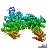 EMDB-4723: 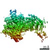 EMDB-4724: 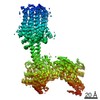 EMDB-4725: 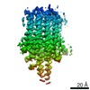 EMDB-4726: |
| Chemicals |  ChemComp-GSP:  ChemComp-MG:  ChemComp-ONM:  ChemComp-FOK:  ChemComp-MN: |
| Source |
|
 Keywords Keywords | MEMBRANE PROTEIN / adenylyl cyclase / G protein / occluded state / MANT-GTP / forskolin |