 Movie
Movie Controller


+ Open data

- Basic information
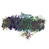
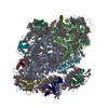
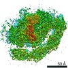
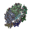
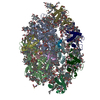
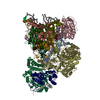
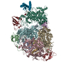
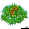
| Entry | Database: PDB / ID: 7blz | ||||||
|---|---|---|---|---|---|---|---|
| Title | Red alga C.merolae Photosystem I | ||||||
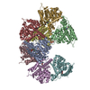
 Components Components |
| ||||||
 Keywords Keywords | PHOTOSYNTHESIS / Red alga C.merolae Photosystem I | ||||||
| Function / homology |  Function and homology information Function and homology informationplastid thylakoid membrane / thylakoid membrane / photosynthesis, light harvesting / photosystem I reaction center / photosystem I / photosynthetic electron transport in photosystem I / photosystem I / chlorophyll binding / chloroplast thylakoid membrane / photosynthesis ...plastid thylakoid membrane / thylakoid membrane / photosynthesis, light harvesting / photosystem I reaction center / photosystem I / photosynthetic electron transport in photosystem I / photosystem I / chlorophyll binding / chloroplast thylakoid membrane / photosynthesis / chloroplast / 4 iron, 4 sulfur cluster binding / electron transfer activity / oxidoreductase activity / magnesium ion binding / membrane / metal ion binding Similarity search - Function | ||||||
| Biological species |  Cyanidioschyzon merolae (eukaryote) Cyanidioschyzon merolae (eukaryote) | ||||||
| Method | ELECTRON MICROSCOPY / single particle reconstruction / cryo EM / Resolution: 3.1 Å | ||||||
 Authors Authors | Nelson, N. / Klaiman, D. / Hippler, M. | ||||||
| Funding support |  Israel, 1items Israel, 1items
| ||||||
 Citation Citation | Journal: To Be Published Title: Red alga C.merolae Photosystem I Authors: Nelson, N. / Klaiman, D. / Hippler, M. | ||||||
| History |
|
- Structure visualization
| Movie |
Movie viewer |
|---|---|
| Structure viewer | Molecule: MolmilJmol/JSmol |
- Downloads & links
-Download
| PDBx/mmCIF format | 7blz.cif.gz | 1.4 MB | Display | PDBx/mmCIF format |
|---|---|---|---|---|
| PDB format | pdb7blz.ent.gz | 1.3 MB | Display | PDB format |
| PDBx/mmJSON format | 7blz.json.gz | Tree view | PDBx/mmJSON format | |
| Others |  Other downloads Other downloads |
-Validation report
| Arichive directory | https://data.pdbj.org/pub/pdb/validation_reports/bl/7blzftp://data.pdbj.org/pub/pdb/validation_reports/bl/7blz | HTTPS FTP |
|---|
-Related structure data
| Related structure data | 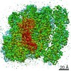 12228MC M: map data used to model this data C: citing same article ( |
|---|---|
| Similar structure data |
-Links
 PDBj
PDBj


- Assembly
| Deposited unit | 
|
|---|---|
| 1 |
|
-Components
-Protein , 6 types, 6 molecules 123FKO
| #1: Protein | Mass: 19551.574 Da / Num. of mol.: 1 / Source method: isolated from a natural source Source: (natural) Cyanidioschyzon merolae (strain 10D) (eukaryote)Strain: 10D / References: UniProt: M1VKK5 |
|---|---|
| #2: Protein | Mass: 19796.850 Da / Num. of mol.: 1 / Source method: isolated from a natural source Source: (natural) Cyanidioschyzon merolae (strain 10D) (eukaryote)Strain: 10D / References: UniProt: M1UU36 |
| #3: Protein | Mass: 19041.045 Da / Num. of mol.: 1 / Source method: isolated from a natural source Source: (natural) Cyanidioschyzon merolae (strain 10D) (eukaryote)Strain: 10D |
| #9: Protein | Mass: 17997.396 Da / Num. of mol.: 1 / Source method: isolated from a natural source Source: (natural) Cyanidioschyzon merolae (strain 10D) (eukaryote)Strain: 10D / References: UniProt: A0A5P9RU83 |
| #12: Protein | Mass: 5608.732 Da / Num. of mol.: 1 / Source method: isolated from a natural source Source: (natural) Cyanidioschyzon merolae (strain 10D) (eukaryote)Strain: 10D / References: UniProt: Q85G51 |
| #15: Protein | Mass: 10485.055 Da / Num. of mol.: 1 / Source method: isolated from a natural source Source: (natural) Cyanidioschyzon merolae (strain 10D) (eukaryote)Strain: 10D / References: UniProt: M1VFJ4 |
-Photosystem I P700 chlorophyll a apoprotein ... , 3 types, 3 molecules ABD
| #4: Protein | Mass: 82215.789 Da / Num. of mol.: 1 / Source method: isolated from a natural source Source: (natural) Cyanidioschyzon merolae (strain 10D) (eukaryote)Strain: 10D / References: UniProt: Q85FY7, photosystem I |
|---|---|
| #5: Protein | Mass: 81976.656 Da / Num. of mol.: 1 / Source method: isolated from a natural source Source: (natural) Cyanidioschyzon merolae (strain 10D) (eukaryote)Strain: 10D / References: UniProt: Q85FY6, photosystem I |
| #7: Protein | Mass: 15567.736 Da / Num. of mol.: 1 / Source method: isolated from a natural source Source: (natural) Cyanidioschyzon merolae (strain 10D) (eukaryote)Strain: 10D / References: UniProt: Q85FY0 |
-Photosystem I iron-sulfur ... , 2 types, 2 molecules CE
| #6: Protein | Mass: 8691.076 Da / Num. of mol.: 1 / Source method: isolated from a natural source Source: (natural) Cyanidioschyzon merolae (strain 10D) (eukaryote)Strain: 10D / References: UniProt: Q85G47, photosystem I |
|---|---|
| #8: Protein | Mass: 6985.040 Da / Num. of mol.: 1 / Source method: isolated from a natural source Source: (natural) Cyanidioschyzon merolae (strain 10D) (eukaryote)Strain: 10D / References: UniProt: Q85FZ1 |
-Photosystem I reaction center subunit ... , 4 types, 4 molecules IJLM
| #10: Protein/peptide | Mass: 3276.988 Da / Num. of mol.: 1 / Source method: isolated from a natural source Source: (natural) Cyanidioschyzon merolae (strain 10D) (eukaryote)Strain: 10D / References: UniProt: Q85FQ6 |
|---|---|
| #11: Protein/peptide | Mass: 4410.245 Da / Num. of mol.: 1 / Source method: isolated from a natural source Source: (natural) Cyanidioschyzon merolae (strain 10D) (eukaryote)Strain: 10D / References: UniProt: A0A5P9RTH6 |
| #13: Protein | Mass: 14690.970 Da / Num. of mol.: 1 / Source method: isolated from a natural source Source: (natural) Cyanidioschyzon merolae (strain 10D) (eukaryote)Strain: 10D / References: UniProt: Q85FP8 |
| #14: Protein/peptide | Mass: 2937.604 Da / Num. of mol.: 1 / Source method: isolated from a natural source Source: (natural) Cyanidioschyzon merolae (strain 10D) (eukaryote)Strain: 10D / References: UniProt: Q85G73 |
-Sugars , 2 types, 5 molecules 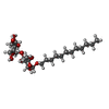

| #24: Sugar |  Type: D-saccharide / Mass: 510.615 Da / Num. of mol.: 2 / Source method: obtained synthetically / Formula: C24H46O11 / Comment: detergent*YM Type: D-saccharide / Mass: 510.615 Da / Num. of mol.: 2 / Source method: obtained synthetically / Formula: C24H46O11 / Comment: detergent*YM#31: Sugar |  Type: saccharide / Mass: 949.299 Da / Num. of mol.: 3 / Source method: obtained synthetically / Formula: C51H96O15 Type: saccharide / Mass: 949.299 Da / Num. of mol.: 3 / Source method: obtained synthetically / Formula: C51H96O15 |
|---|
-Non-polymers , 16 types, 450 molecules 
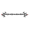


| #16: Chemical | ChemComp-CLA /  Mass: 893.489 Da / Num. of mol.: 132 / Source method: obtained synthetically / Formula: C55H72MgN4O5 Mass: 893.489 Da / Num. of mol.: 132 / Source method: obtained synthetically / Formula: C55H72MgN4O5#17: Chemical | ChemComp-C7Z / (  Mass: 568.871 Da / Num. of mol.: 13 / Source method: obtained synthetically / Formula: C40H56O2 Mass: 568.871 Da / Num. of mol.: 13 / Source method: obtained synthetically / Formula: C40H56O2#18: Chemical | ChemComp-RRX / (  Mass: 552.872 Da / Num. of mol.: 5 / Source method: obtained synthetically / Formula: C40H56O Mass: 552.872 Da / Num. of mol.: 5 / Source method: obtained synthetically / Formula: C40H56O#19: Chemical | ChemComp-LHG /  Mass: 722.970 Da / Num. of mol.: 6 / Source method: obtained synthetically / Formula: C38H75O10P / Comment: phospholipid*YM Mass: 722.970 Da / Num. of mol.: 6 / Source method: obtained synthetically / Formula: C38H75O10P / Comment: phospholipid*YM#20: Chemical |  Mass: 396.648 Da / Num. of mol.: 3 / Source method: obtained synthetically / Formula: C28H44O Mass: 396.648 Da / Num. of mol.: 3 / Source method: obtained synthetically / Formula: C28H44O#21: Chemical | ChemComp-BCR /  Mass: 536.873 Da / Num. of mol.: 22 / Source method: obtained synthetically / Formula: C40H56 Mass: 536.873 Da / Num. of mol.: 22 / Source method: obtained synthetically / Formula: C40H56#22: Chemical |  Mass: 751.023 Da / Num. of mol.: 2 / Source method: obtained synthetically / Formula: C40H79O10P / Comment: phospholipid*YM Mass: 751.023 Da / Num. of mol.: 2 / Source method: obtained synthetically / Formula: C40H79O10P / Comment: phospholipid*YM#23: Chemical |  Mass: 625.018 Da / Num. of mol.: 2 / Source method: obtained synthetically / Formula: C39H76O5 Mass: 625.018 Da / Num. of mol.: 2 / Source method: obtained synthetically / Formula: C39H76O5#25: Chemical | ChemComp-PTY /  Mass: 734.039 Da / Num. of mol.: 4 / Source method: obtained synthetically / Formula: C40H80NO8P / Comment: phospholipid*YM Mass: 734.039 Da / Num. of mol.: 4 / Source method: obtained synthetically / Formula: C40H80NO8P / Comment: phospholipid*YM#26: Chemical | ChemComp-CL0 / |  Mass: 893.489 Da / Num. of mol.: 1 / Source method: obtained synthetically / Formula: C55H72MgN4O5 Mass: 893.489 Da / Num. of mol.: 1 / Source method: obtained synthetically / Formula: C55H72MgN4O5#27: Chemical |  Mass: 450.696 Da / Num. of mol.: 2 / Source method: obtained synthetically / Formula: C31H46O2 Mass: 450.696 Da / Num. of mol.: 2 / Source method: obtained synthetically / Formula: C31H46O2#28: Chemical |  Mass: 351.640 Da / Num. of mol.: 3 / Source method: obtained synthetically / Formula: Fe4S4 Mass: 351.640 Da / Num. of mol.: 3 / Source method: obtained synthetically / Formula: Fe4S4#29: Chemical |  Mass: 704.998 Da / Num. of mol.: 3 / Source method: obtained synthetically / Formula: C39H77O8P Mass: 704.998 Da / Num. of mol.: 3 / Source method: obtained synthetically / Formula: C39H77O8P#30: Chemical | ChemComp-T7X / |  Mass: 887.128 Da / Num. of mol.: 1 / Source method: obtained synthetically / Formula: C47H83O13P Mass: 887.128 Da / Num. of mol.: 1 / Source method: obtained synthetically / Formula: C47H83O13P#32: Chemical |  Mass: 40.078 Da / Num. of mol.: 2 / Source method: isolated from a natural source / Formula: Ca Mass: 40.078 Da / Num. of mol.: 2 / Source method: isolated from a natural source / Formula: Ca#33: Water | ChemComp-HOH / | Mass: 18.015 Da / Num. of mol.: 249 / Source method: isolated from a natural source / Formula: H2O |
|---|
-Details
| Has ligand of interest | N |
|---|---|
| Has protein modification | Y |
-Experimental details
-Experiment
| Experiment | Method: ELECTRON MICROSCOPY |
|---|---|
| EM experiment | Aggregation state: PARTICLE / 3D reconstruction method: single particle reconstruction |
- Sample preparation
| Component | Name: Photosystem I / Type: COMPLEX / Entity ID: #1-#15 / Source: NATURAL |
|---|---|
| Source (natural) | Organism: Cyanidioschyzon merolae strain 10D (eukaryote) |
| Buffer solution | pH: 8 |
| Specimen | Embedding applied: NO / Shadowing applied: NO / Staining applied: NO / Vitrification applied: YES |
| Specimen support | Grid material: COPPER / Grid type: Quantifoil R1.2/1.3 |
| Vitrification | Cryogen name: ETHANE |
- Electron microscopy imaging
| Experimental equipment |  Model: Titan Krios / Image courtesy: FEI Company |
|---|---|
| Microscopy | Model: FEI TITAN KRIOS |
| Electron gun | Electron source:  FIELD EMISSION GUN / Accelerating voltage: 300 kV / Illumination mode: FLOOD BEAM FIELD EMISSION GUN / Accelerating voltage: 300 kV / Illumination mode: FLOOD BEAM |
| Electron lens | Mode: BRIGHT FIELD |
| Image recording | Electron dose: 40 e/Å2 / Film or detector model: GATAN K3 BIOQUANTUM (6k x 4k) / Num. of real images: 7220 |
- Processing
| Software | Name: PHENIX / Version: 1.19_4092: / Classification: refinement | ||||||||||||||||||||||||||||||||
|---|---|---|---|---|---|---|---|---|---|---|---|---|---|---|---|---|---|---|---|---|---|---|---|---|---|---|---|---|---|---|---|---|---|
| EM software |
| ||||||||||||||||||||||||||||||||
| CTF correction | Type: NONE | ||||||||||||||||||||||||||||||||
| Particle selection | Num. of particles selected: 1198564 | ||||||||||||||||||||||||||||||||
| 3D reconstruction | Resolution: 3.1 Å / Resolution method: FSC 0.143 CUT-OFF / Num. of particles: 128943 / Num. of class averages: 8 / Symmetry type: POINT | ||||||||||||||||||||||||||||||||
| Atomic model building | Protocol: RIGID BODY FIT / Space: REAL | ||||||||||||||||||||||||||||||||
| Refinement | Cross valid method: NONE Stereochemistry target values: GeoStd + Monomer Library + CDL v1.2 | ||||||||||||||||||||||||||||||||
| Displacement parameters | Biso mean: 35.12 Å2 | ||||||||||||||||||||||||||||||||
| Refine LS restraints |
|
