+ Open data
- Basic information
| Entry | Database: EMDB / ID: EMD-12228 | |||||||||
|---|---|---|---|---|---|---|---|---|---|---|
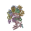
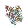
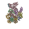
| Title | Red alga C.merolae Photosystem I | |||||||||
 Map data Map data | ||||||||||
 Sample Sample |
| |||||||||
 Keywords Keywords | Red alga C.merolae Photosystem I / PHOTOSYNTHESIS | |||||||||
| Function / homology |  Function and homology information Function and homology informationplastid thylakoid membrane / thylakoid membrane / photosynthesis, light harvesting / photosystem I reaction center / photosystem I / photosynthetic electron transport in photosystem I / photosystem I / chlorophyll binding / chloroplast thylakoid membrane / photosynthesis ...plastid thylakoid membrane / thylakoid membrane / photosynthesis, light harvesting / photosystem I reaction center / photosystem I / photosynthetic electron transport in photosystem I / photosystem I / chlorophyll binding / chloroplast thylakoid membrane / photosynthesis / chloroplast / 4 iron, 4 sulfur cluster binding / oxidoreductase activity / electron transfer activity / magnesium ion binding / metal ion binding / membrane Similarity search - Function | |||||||||
| Biological species |  Cyanidioschyzon merolae strain 10D (eukaryote) / Cyanidioschyzon merolae strain 10D (eukaryote) /  Cyanidioschyzon merolae (strain 10D) (eukaryote) Cyanidioschyzon merolae (strain 10D) (eukaryote) | |||||||||
| Method | single particle reconstruction / cryo EM / Resolution: 3.1 Å | |||||||||
 Authors Authors | Nelson N / Klaiman D | |||||||||
| Funding support |  Israel, 1 items Israel, 1 items
| |||||||||
 Citation Citation |  Journal: To Be Published Journal: To Be PublishedTitle: Red alga C.merolae Photosystem I Authors: Nelson N / Klaiman D / Hippler M | |||||||||
| History |
|
- Structure visualization
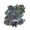
| Movie |
 Movie viewer Movie viewer |
|---|---|
| Structure viewer | EM map:  SurfView SurfView Molmil Molmil Jmol/JSmol Jmol/JSmol |
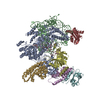
| Supplemental images |
- Downloads & links
-EMDB archive
| Map data |  emd_12228.map.gz emd_12228.map.gz | 96.5 MB |  EMDB map data format EMDB map data format | |
|---|---|---|---|---|
| Header (meta data) |  emd-12228-v30.xml emd-12228-v30.xml emd-12228.xml emd-12228.xml | 35.8 KB 35.8 KB | Display Display |  EMDB header EMDB header |
| FSC (resolution estimation) |  emd_12228_fsc.xml emd_12228_fsc.xml | 10.6 KB | Display |  FSC data file FSC data file |
| Images |  emd_12228.png emd_12228.png | 253.4 KB | ||
| Filedesc metadata |  emd-12228.cif.gz emd-12228.cif.gz | 8.5 KB | ||
| Others |  emd_12228_half_map_1.map.gz emd_12228_half_map_1.map.gz emd_12228_half_map_2.map.gz emd_12228_half_map_2.map.gz | 80.8 MB 80.8 MB | ||
| Archive directory |  http://ftp.pdbj.org/pub/emdb/structures/EMD-12228 http://ftp.pdbj.org/pub/emdb/structures/EMD-12228 ftp://ftp.pdbj.org/pub/emdb/structures/EMD-12228 ftp://ftp.pdbj.org/pub/emdb/structures/EMD-12228 | HTTPS FTP |
-Validation report
| Summary document |  emd_12228_validation.pdf.gz emd_12228_validation.pdf.gz | 1 MB | Display |  EMDB validaton report EMDB validaton report |
|---|---|---|---|---|
| Full document |  emd_12228_full_validation.pdf.gz emd_12228_full_validation.pdf.gz | 1023.7 KB | Display | |
| Data in XML |  emd_12228_validation.xml.gz emd_12228_validation.xml.gz | 17.9 KB | Display | |
| Data in CIF |  emd_12228_validation.cif.gz emd_12228_validation.cif.gz | 23.6 KB | Display | |
| Arichive directory |  https://ftp.pdbj.org/pub/emdb/validation_reports/EMD-12228 https://ftp.pdbj.org/pub/emdb/validation_reports/EMD-12228 ftp://ftp.pdbj.org/pub/emdb/validation_reports/EMD-12228 ftp://ftp.pdbj.org/pub/emdb/validation_reports/EMD-12228 | HTTPS FTP |
-Related structure data
| Related structure data |  7blzMC M: atomic model generated by this map C: citing same article ( |
|---|---|
| Similar structure data |
- Links
Links
| EMDB pages |  EMDB (EBI/PDBe) / EMDB (EBI/PDBe) /  EMDataResource EMDataResource |
|---|---|
| Related items in Molecule of the Month |
- Map
Map
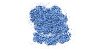
| File |  Download / File: emd_12228.map.gz / Format: CCP4 / Size: 103 MB / Type: IMAGE STORED AS FLOATING POINT NUMBER (4 BYTES) Download / File: emd_12228.map.gz / Format: CCP4 / Size: 103 MB / Type: IMAGE STORED AS FLOATING POINT NUMBER (4 BYTES) | ||||||||||||||||||||||||||||||||||||||||||||||||||||||||||||
|---|---|---|---|---|---|---|---|---|---|---|---|---|---|---|---|---|---|---|---|---|---|---|---|---|---|---|---|---|---|---|---|---|---|---|---|---|---|---|---|---|---|---|---|---|---|---|---|---|---|---|---|---|---|---|---|---|---|---|---|---|---|
| Projections & slices | Image control
Images are generated by Spider. | ||||||||||||||||||||||||||||||||||||||||||||||||||||||||||||
| Voxel size | X=Y=Z: 1.052 Å | ||||||||||||||||||||||||||||||||||||||||||||||||||||||||||||
| Density |
| ||||||||||||||||||||||||||||||||||||||||||||||||||||||||||||
| Symmetry | Space group: 1 | ||||||||||||||||||||||||||||||||||||||||||||||||||||||||||||
| Details | EMDB XML:
CCP4 map header:
| ||||||||||||||||||||||||||||||||||||||||||||||||||||||||||||
-Supplemental data
-Half map: #2
| File | emd_12228_half_map_1.map | ||||||||||||
|---|---|---|---|---|---|---|---|---|---|---|---|---|---|
| Projections & Slices |
| ||||||||||||
| Density Histograms |
-Half map: #1
| File | emd_12228_half_map_2.map | ||||||||||||
|---|---|---|---|---|---|---|---|---|---|---|---|---|---|
| Projections & Slices |
| ||||||||||||
| Density Histograms |
- Sample components
+Entire : Photosystem I
+Supramolecule #1: Photosystem I
+Macromolecule #1: Similar to light harvesting protein
+Macromolecule #2: Similar to chlorophyll a/b-binding protein, CP24
+Macromolecule #3: Lhcr3
+Macromolecule #4: Photosystem I P700 chlorophyll a apoprotein A1
+Macromolecule #5: Photosystem I P700 chlorophyll a apoprotein A2
+Macromolecule #6: Photosystem I iron-sulfur center
+Macromolecule #7: Photosystem I p700 chlorophyll A apoprotein A2
+Macromolecule #8: Photosystem I iron-sulfur center subunit VII
+Macromolecule #9: PSI-F
+Macromolecule #10: Photosystem I reaction center subunit VIII
+Macromolecule #11: Photosystem I reaction center subunit IX
+Macromolecule #12: PSI-K
+Macromolecule #13: Photosystem I reaction center subunit XI
+Macromolecule #14: Photosystem I reaction center subunit XII
+Macromolecule #15: PsaO
+Macromolecule #16: CHLOROPHYLL A
+Macromolecule #17: (1~{S})-3,5,5-trimethyl-4-[(1~{E},3~{E},5~{E},7~{E},9~{E},11~{E},...
+Macromolecule #18: (3R)-beta,beta-caroten-3-ol
+Macromolecule #19: 1,2-DIPALMITOYL-PHOSPHATIDYL-GLYCEROLE
+Macromolecule #20: ERGOSTEROL
+Macromolecule #21: BETA-CAROTENE
+Macromolecule #22: (1S)-2-{[{[(2R)-2,3-DIHYDROXYPROPYL]OXY}(HYDROXY)PHOSPHORYL]OXY}-...
+Macromolecule #23: DIACYL GLYCEROL
+Macromolecule #24: DODECYL-ALPHA-D-MALTOSIDE
+Macromolecule #25: PHOSPHATIDYLETHANOLAMINE
+Macromolecule #26: CHLOROPHYLL A ISOMER
+Macromolecule #27: PHYLLOQUINONE
+Macromolecule #28: IRON/SULFUR CLUSTER
+Macromolecule #29: 1,2-DIACYL-GLYCEROL-3-SN-PHOSPHATE
+Macromolecule #30: Phosphatidylinositol
+Macromolecule #31: DIGALACTOSYL DIACYL GLYCEROL (DGDG)
+Macromolecule #32: CALCIUM ION
+Macromolecule #33: water
-Experimental details
-Structure determination
| Method | cryo EM |
|---|---|
 Processing Processing | single particle reconstruction |
| Aggregation state | particle |
- Sample preparation
| Buffer | pH: 8 |
|---|---|
| Grid | Model: Quantifoil R1.2/1.3 / Material: COPPER |
| Vitrification | Cryogen name: ETHANE |
- Electron microscopy
| Microscope | FEI TITAN KRIOS |
|---|---|
| Image recording | Film or detector model: GATAN K3 BIOQUANTUM (6k x 4k) / Number real images: 7220 / Average electron dose: 40.0 e/Å2 |
| Electron beam | Acceleration voltage: 300 kV / Electron source:  FIELD EMISSION GUN FIELD EMISSION GUN |
| Electron optics | Illumination mode: FLOOD BEAM / Imaging mode: BRIGHT FIELD |
| Experimental equipment |  Model: Titan Krios / Image courtesy: FEI Company |
+ Image processing
-Atomic model buiding 1
| Refinement | Space: REAL / Protocol: RIGID BODY FIT |
|---|---|
| Output model |  PDB-7blz: |
 Movie
Movie Controller






 Z (Sec.)
 Y (Row.)
 X (Col.)


