 Movie
Movie Controller

[English] 日本語
 Yorodumi
- PDB-3q1r: Crystal structure of a bacterial RNase P holoenzyme in complex wi... -
+ Open data

- Basic information
| Entry | Database: PDB / ID: 3q1r | |||||||||
|---|---|---|---|---|---|---|---|---|---|---|
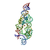
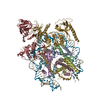
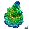
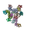
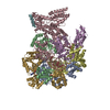
| Title | Crystal structure of a bacterial RNase P holoenzyme in complex with TRNA and in the presence of 5' leader | |||||||||
 Components Components |
| |||||||||
 Keywords Keywords | HYDROLASE/RNA / RNASE P / RIBOZYME / RIBONUCLEASE P / TRNA / PRE-TRNA / TETRALOOP-RECEPTOR / RIBOSE ZIPPER / A-MINOR INTERACTION / BASE STACKING / INTERMOLECULAR BASE PAIRS / INTERMOLECULAR RNA-RNA CONTACTS / RNP / RIBONUCLEOPROTEIN COMPLEX / ENZYME-PRODUCT COMPLEX / METALLOENZYME / RNA-METAL INTERACTIONS / SHAPE COMPLEMENTARITY / SUBSTRATE RECOGNITION / ENDONUCLEASE / HYDROLASE-RNA COMPLEX | |||||||||
| Function / homology |  Function and homology information Function and homology informationtRNA 3'-end processing / 3'-tRNA processing endoribonuclease activity / ribonuclease P complex / ribonuclease P / ribonuclease P activity / tRNA 5'-leader removal / tRNA binding Similarity search - Function | |||||||||
| Biological species |   Thermotoga maritima (bacteria) Thermotoga maritima (bacteria) | |||||||||
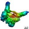
| Method |  X-RAY DIFFRACTION / SYNCHROTRON / MIRAS / Resolution: 4.21 Å X-RAY DIFFRACTION / SYNCHROTRON / MIRAS / Resolution: 4.21 Å | |||||||||
 Authors Authors | Reiter, N.J. / Ostermanm, A. / Torres-Larios, A. / Swinger, K.K. / Pan, T. / Mondragon, A. | |||||||||
 Citation Citation | Journal: Nature / Year: 2010 Title: Structure of a Bacterial Ribonuclease P Holoenzyme in Complex with tRNA. Authors: Reiter, N.J. / Osterman, A. / Torres-Larios, A. / Swinger, K.K. / Pan, T. / Mondragon, A. | |||||||||
| History |
| |||||||||
| Remark 650 | HELIX DETERMINATION METHOD: AUTHOR |
- Structure visualization
| Structure viewer | Molecule: MolmilJmol/JSmol |
|---|
- Downloads & links
-Download
| PDBx/mmCIF format | 3q1r.cif.gz | 240.5 KB | Display | PDBx/mmCIF format |
|---|---|---|---|---|
| PDB format | pdb3q1r.ent.gz | 175.4 KB | Display | PDB format |
| PDBx/mmJSON format | 3q1r.json.gz | Tree view | PDBx/mmJSON format | |
| Others |  Other downloads Other downloads |
-Validation report
| Arichive directory | https://data.pdbj.org/pub/pdb/validation_reports/q1/3q1rftp://data.pdbj.org/pub/pdb/validation_reports/q1/3q1r | HTTPS FTP |
|---|
-Related structure data
| Related structure data |  3q1qC 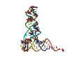 1ehzS 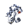 1nz0S 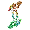 2a2eS 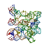 2a64S S: Starting model for refinement C: citing same article ( |
|---|---|
| Similar structure data |


-Links
 PDBj
PDBj


- Assembly
| Deposited unit | 
| ||||||||
|---|---|---|---|---|---|---|---|---|---|
| 1 |
| ||||||||
| Unit cell |
|
-Components
| #1: RNA chain | Mass: 112622.805 Da / Num. of mol.: 1 / Source method: obtained synthetically / Details: THE RNA WAS PREPARED BY IN VITRO TRANSCRIPTION |
|---|---|
| #2: Protein | Mass: 14363.003 Da / Num. of mol.: 1 Source method: isolated from a genetically manipulated source Source: (gene. exp.) Thermotoga maritima (bacteria) / Gene: rnpA, RNPA OR TM1463, RNPB, TM_1463 / Plasmid: PGEX4T-2 / Production host: |
| #3: RNA chain | Mass: 27658.418 Da / Num. of mol.: 1 / Source method: obtained synthetically / Details: THE RNA WAS PREPARED BY IN VITRO TRANSCRIPTION |
| #4: RNA chain | Mass: 2260.419 Da / Num. of mol.: 1 / Source method: obtained synthetically |
| #5: Chemical | ChemComp-MG /   Mass: 24.305 Da / Num. of mol.: 5 / Source method: obtained synthetically / Formula: Mg Mass: 24.305 Da / Num. of mol.: 5 / Source method: obtained synthetically / Formula: Mg |
-Experimental details
-Experiment
| Experiment | Method: X-RAY DIFFRACTION / Number of used crystals: 1 |
|---|
- Sample preparation
| Crystal | Density Matthews: 4.95 Å3/Da / Density % sol: 75.14 % Description: DUE TO THE ANISOTROPIC NATURE OF THE DIFFRACTION, THE DATA WERE TREATED IN TWO DIFFERENT WAYS: 1) BY APPLYING AN ANISOTROPIC CORRECTION TO THE DATA (ANISOTROPIC), AND 2) WITHOUT ANY ...Description: DUE TO THE ANISOTROPIC NATURE OF THE DIFFRACTION, THE DATA WERE TREATED IN TWO DIFFERENT WAYS: 1) BY APPLYING AN ANISOTROPIC CORRECTION TO THE DATA (ANISOTROPIC), AND 2) WITHOUT ANY ANISOTROPY CORRECTION (ISOTROPIC). WHILE COMPLETENESS OF THE HIGHEST RESOLUTION SHELL MAY APPEAR OF POOR QUALITY IN THE ANISOTROPIC CASE, THE DATA COLLECTION STATISTICS AND RWORK AND RFREE REFINEMENT STATISTICS WERE ACTUALLY BETTER FOR THE CARVED (ANISOTROPIC) DATA SET THAN FOR THE ISOTROPIC CASE. FOR THIS REASON, THE REPORTED 3Q1R STRUCTURE AND HEADER REMARKS REFLECT THE CARVED, ANISOTROPICALLY CORRECTED DATA. |
|---|---|
| Crystal grow | Temperature: 303 K / Method: vapor diffusion, hanging drop / pH: 6 Details: 1.8M LiSO4, 0.5M sodium cacolydate, pH 6.0, VAPOR DIFFUSION, HANGING DROP, temperature 303K |
-Data collection
| Diffraction | Mean temperature: 100 K |
|---|---|
| Diffraction source | Source: SYNCHROTRON / Site: APS  / Beamline: 21-ID-F / Wavelength: 0.97872 / Beamline: 21-ID-F / Wavelength: 0.97872 |
| Detector | Type: RAYONIX MX-225 / Detector: CCD / Date: Feb 10, 2010 / Details: MIRRORS |
| Radiation | Monochromator: DIAMOND LAUE / Protocol: SINGLE WAVELENGTH / Monochromatic (M) / Laue (L): M / Scattering type: x-ray |
| Radiation wavelength | Wavelength: 0.97872 Å / Relative weight: 1 |
| Reflection | Resolution: 4.2→30 Å / Num. obs: 17674 / % possible obs: 76.4 % / Redundancy: 5.7 % / Biso Wilson estimate: 153.42 Å2 / Rmerge(I) obs: 0.081 / Rsym value: 0.099 / Net I/σ(I): 10.7 |
| Reflection shell | Resolution: 4.2→4.39 Å / Redundancy: 3.7 % / Rmerge(I) obs: 0.463 / Mean I/σ(I) obs: 2.4 / Rsym value: 0.577 / % possible all: 7.8 |
- Processing
| Software |
| ||||||||||||||||||||||||||||||||||||||||
|---|---|---|---|---|---|---|---|---|---|---|---|---|---|---|---|---|---|---|---|---|---|---|---|---|---|---|---|---|---|---|---|---|---|---|---|---|---|---|---|---|---|
| Refinement | Method to determine structure: MIRAS Starting model: 2A2E, 2A64, 1NZ0, 1EHZ Resolution: 4.21→29.63 Å / Cor.coef. Fo:Fc: 0.895 / Cor.coef. Fo:Fc free: 0.882 / Cross valid method: THROUGHOUT / σ(F): 0
| ||||||||||||||||||||||||||||||||||||||||
| Displacement parameters | Biso mean: 80.04 Å2
| ||||||||||||||||||||||||||||||||||||||||
| Refine analyze | Luzzati coordinate error obs: 1.03 Å | ||||||||||||||||||||||||||||||||||||||||
| Refinement step | Cycle: LAST / Resolution: 4.21→29.63 Å
| ||||||||||||||||||||||||||||||||||||||||
| Refine LS restraints |
| ||||||||||||||||||||||||||||||||||||||||
| LS refinement shell | Resolution: 4.21→4.46 Å / Total num. of bins used: 9
|
