 Movie
Movie Controller

[English] 日本語
 Yorodumi
- PDB-1nbs: Crystal structure of the specificity domain of Ribonuclease P RNA -
+ Open data

- Basic information
| Entry | Database: PDB / ID: 1nbs | ||||||
|---|---|---|---|---|---|---|---|
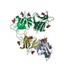
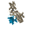
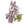
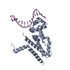
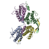
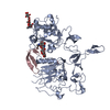
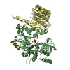
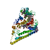
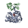
| Title | Crystal structure of the specificity domain of Ribonuclease P RNA | ||||||
 Components Components | RIBONUCLEASE P RNA | ||||||
 Keywords Keywords | RNA / Ribonuclease P RNA / P RNA / S-domain | ||||||
| Function / homology | LEAD (II) ION / : / RNA / RNA (> 10) / RNA (> 100) Function and homology information Function and homology information | ||||||
| Biological species |  | ||||||
| Method |  X-RAY DIFFRACTION / SYNCHROTRON / MAD / Resolution: 3.15 Å X-RAY DIFFRACTION / SYNCHROTRON / MAD / Resolution: 3.15 Å | ||||||
 Authors Authors | Krasilnikov, A.S. / Yang, X. / Pan, T. / Mondragon, A. | ||||||
 Citation Citation | Journal: Nature / Year: 2003 Title: Crystal structure of the specificity domain of Ribonuclease P Authors: Krasilnikov, A.S. / Yang, X. / Pan, T. / Mondragon, A. | ||||||
| History |
| ||||||
| Remark 999 | SEQUENCE Base A240 is a transcriptional artifact. |
- Structure visualization
| Structure viewer | Molecule: MolmilJmol/JSmol |
|---|
- Downloads & links
-Download
| PDBx/mmCIF format | 1nbs.cif.gz | 158.9 KB | Display | PDBx/mmCIF format |
|---|---|---|---|---|
| PDB format | pdb1nbs.ent.gz | 121.5 KB | Display | PDB format |
| PDBx/mmJSON format | 1nbs.json.gz | Tree view | PDBx/mmJSON format | |
| Others |  Other downloads Other downloads |
-Validation report
| Arichive directory | https://data.pdbj.org/pub/pdb/validation_reports/nb/1nbsftp://data.pdbj.org/pub/pdb/validation_reports/nb/1nbs | HTTPS FTP |
|---|
-Related structure data
| Related structure data | |
|---|---|
| Similar structure data |


-Links
 PDBj
PDBj


- Assembly
| Deposited unit | 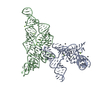
| ||||||||
|---|---|---|---|---|---|---|---|---|---|
| 1 | 
| ||||||||
| 2 | 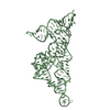
| ||||||||
| Unit cell |
|
-Components
| #1: RNA chain | Mass: 50209.945 Da / Num. of mol.: 2 / Fragment: Specificity-domain, S-domain / Mutation: U86G, A239C, U89A Source method: isolated from a genetically manipulated source Source: (gene. exp.) Description: The RNA was produced by vitro transcription with T7 polymerase from a pUC19-based template which contained cloned sequence of the S-domain. Plasmid: pUC19 / Production host: #2: Chemical | ChemComp-MG /   Mass: 24.305 Da / Num. of mol.: 12 / Source method: obtained synthetically / Formula: Mg Mass: 24.305 Da / Num. of mol.: 12 / Source method: obtained synthetically / Formula: Mg#3: Chemical | ChemComp-PB /   Mass: 207.200 Da / Num. of mol.: 23 / Source method: obtained synthetically / Formula: Pb Mass: 207.200 Da / Num. of mol.: 23 / Source method: obtained synthetically / Formula: Pb |
|---|
-Experimental details
-Experiment
| Experiment | Method: X-RAY DIFFRACTION / Number of used crystals: 4 |
|---|
- Sample preparation
| Crystal | Density Matthews: 3.31 Å3/Da / Density % sol: 62.82 % | |||||||||||||||||||||||||||||||||||||||||||||||||
|---|---|---|---|---|---|---|---|---|---|---|---|---|---|---|---|---|---|---|---|---|---|---|---|---|---|---|---|---|---|---|---|---|---|---|---|---|---|---|---|---|---|---|---|---|---|---|---|---|---|---|
| Crystal grow | Temperature: 303 K / Method: vapor diffusion, hanging drop / pH: 6 Details: isopropanol, strontium chloride, magnesium chloride, spermine, MES, pH 6.0, VAPOR DIFFUSION, HANGING DROP, temperature 303K | |||||||||||||||||||||||||||||||||||||||||||||||||
| Crystal grow | *PLUS Temperature: 30 ℃ | |||||||||||||||||||||||||||||||||||||||||||||||||
| Components of the solutions | *PLUS
|
-Data collection
| Diffraction | Mean temperature: 100 K | |||||||||
|---|---|---|---|---|---|---|---|---|---|---|
| Diffraction source | Source: SYNCHROTRON / Site: APS  / Beamline: 5ID-B / Wavelength: 0.9475,0.9293 / Beamline: 5ID-B / Wavelength: 0.9475,0.9293 | |||||||||
| Detector | Type: MARRESEARCH / Detector: CCD / Date: Apr 23, 2002 / Details: mirrors | |||||||||
| Radiation | Monochromator: Si MIRRORS / Protocol: MAD / Monochromatic (M) / Laue (L): M / Scattering type: x-ray | |||||||||
| Radiation wavelength |
| |||||||||
| Reflection | Resolution: 3.15→33 Å / Num. all: 23276 / Num. obs: 23276 / % possible obs: 98.2 % / Observed criterion σ(F): 0 / Observed criterion σ(I): 0 / Redundancy: 10.1 % / Biso Wilson estimate: 66.3 Å2 / Rsym value: 0.076 / Net I/σ(I): 7.8 | |||||||||
| Reflection shell | Resolution: 3.15→3.24 Å / Redundancy: 2.9 % / Mean I/σ(I) obs: 2.7 / Num. unique all: 1345 / Rsym value: 0.272 / % possible all: 80.9 | |||||||||
| Reflection | *PLUS Lowest resolution: 33 Å / Num. measured all: 231005 / Rmerge(I) obs: 0.076 | |||||||||
| Reflection shell | *PLUS % possible obs: 80.9 % / Rmerge(I) obs: 0.272 |
- Processing
| Software |
| |||||||||||||||||||||||||||||||||||||||||||||||||||||||
|---|---|---|---|---|---|---|---|---|---|---|---|---|---|---|---|---|---|---|---|---|---|---|---|---|---|---|---|---|---|---|---|---|---|---|---|---|---|---|---|---|---|---|---|---|---|---|---|---|---|---|---|---|---|---|---|---|
| Refinement | Method to determine structure: MAD / Resolution: 3.15→33 Å / Cor.coef. Fo:Fc: 0.857 / Cor.coef. Fo:Fc free: 0.847 / Isotropic thermal model: isotropic / Cross valid method: THROUGHOUT / σ(F): 0 / σ(I): 0 / ESU R Free: 0.515 Details: Bases 143-165, 201-212 in molecule A and 121-124 and 240 in molecule B are disordered; only phosphate backbone could be traced for bases 99 and 101 in both molecules. CNS was also used in refinement.
| |||||||||||||||||||||||||||||||||||||||||||||||||||||||
| Solvent computation | Ion probe radii: 0.8 Å / Shrinkage radii: 0.8 Å / VDW probe radii: 1.4 Å / Solvent model: BABINET MODEL WITH MASK | |||||||||||||||||||||||||||||||||||||||||||||||||||||||
| Displacement parameters | Biso mean: 94.249 Å2
| |||||||||||||||||||||||||||||||||||||||||||||||||||||||
| Refine analyze |
| |||||||||||||||||||||||||||||||||||||||||||||||||||||||
| Refinement step | Cycle: LAST / Resolution: 3.15→33 Å
| |||||||||||||||||||||||||||||||||||||||||||||||||||||||
| Refine LS restraints |
| |||||||||||||||||||||||||||||||||||||||||||||||||||||||
| LS refinement shell | Resolution: 3.154→3.235 Å / Total num. of bins used: 20 /
| |||||||||||||||||||||||||||||||||||||||||||||||||||||||
| Software | *PLUS Name: REFMAC / Version: 5 / Classification: refinement | |||||||||||||||||||||||||||||||||||||||||||||||||||||||
| Refinement | *PLUS Lowest resolution: 33 Å / % reflection Rfree: 5 % / Rfactor Rfree: 0.307 / Rfactor Rwork: 0.28 | |||||||||||||||||||||||||||||||||||||||||||||||||||||||
| Solvent computation | *PLUS | |||||||||||||||||||||||||||||||||||||||||||||||||||||||
| Displacement parameters | *PLUS | |||||||||||||||||||||||||||||||||||||||||||||||||||||||
| Refine LS restraints | *PLUS
|
