- EMDB-4883: Structure of a minimal photosystem I from a green alga -
+
Open data
ID or keywords:
Loading...
-
Basic information
Entry
Database: EMDB / ID: EMD-4883
Title
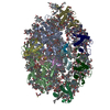
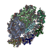
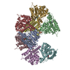
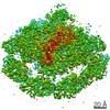
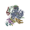
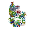
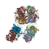
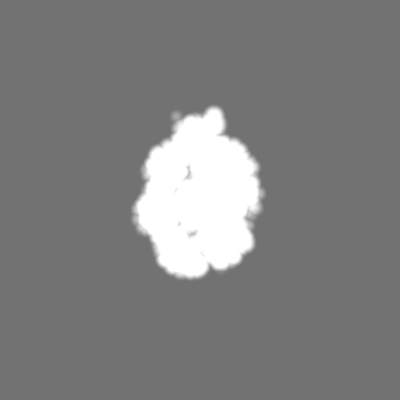
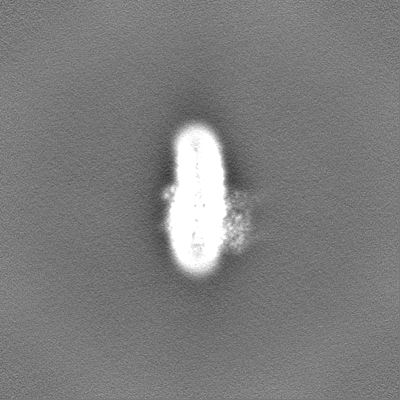
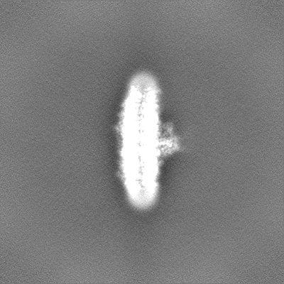
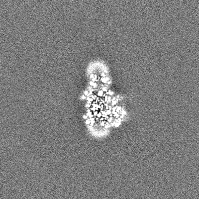
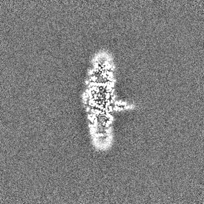
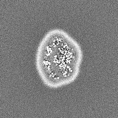
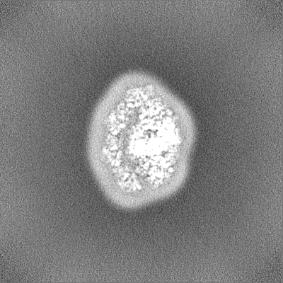
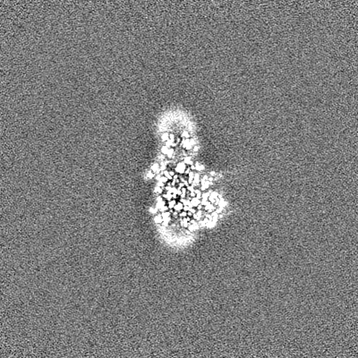
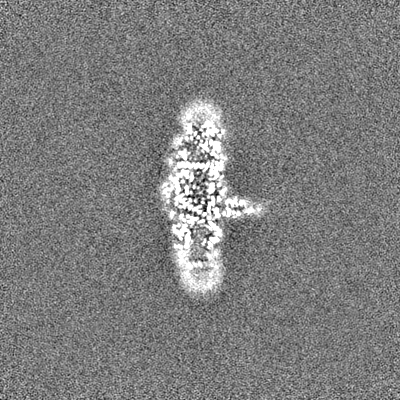
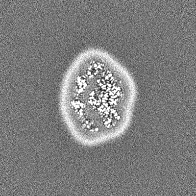
Structure of a minimal photosystem I from a green alga
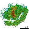
Map data
Sample
Complex: Photosystem I
Protein or peptide: x 11 types
Ligand: x 11 types
Keywords
photosystem I / photosynthesis / green algae
Function / homology
Function and homology information
photosynthesis, light harvesting / photosystem I / photosynthetic electron transport in photosystem I / photosystem I / photosystem II / chlorophyll binding / chloroplast thylakoid membrane / photosynthesis / 4 iron, 4 sulfur cluster binding / electron transfer activity ...photosynthesis, light harvesting / photosystem I / photosynthetic electron transport in photosystem I / photosystem I / photosystem II / chlorophyll binding / chloroplast thylakoid membrane / photosynthesis / 4 iron, 4 sulfur cluster binding / electron transfer activity / oxidoreductase activity / magnesium ion binding / metal ion binding Similarity search - Function
Chlorophyll A-B binding protein, plant and chromista / Chlorophyll A-B binding protein / Chlorophyll A-B binding protein / Photosystem I PsaJ, reaction centre subunit IX superfamily / Photosystem I PsaJ, reaction centre subunit IX / Photosystem I reaction centre subunit IX / PsaJ / Photosystem I PsaA / Photosystem I protein PsaC / Photosystem I PsaB / Photosystem I PsaA/PsaB, conserved site ...Chlorophyll A-B binding protein, plant and chromista / Chlorophyll A-B binding protein / Chlorophyll A-B binding protein / Photosystem I PsaJ, reaction centre subunit IX superfamily / Photosystem I PsaJ, reaction centre subunit IX / Photosystem I reaction centre subunit IX / PsaJ / Photosystem I PsaA / Photosystem I protein PsaC / Photosystem I PsaB / Photosystem I PsaA/PsaB, conserved site / Photosystem I psaA and psaB proteins signature. / : / Photosystem I PsaA/PsaB / Photosystem I PsaA/PsaB superfamily / Photosystem I psaA/psaB protein / 4Fe-4S dicluster domain / 4Fe-4S ferredoxin, iron-sulphur binding, conserved site / 4Fe-4S ferredoxin-type iron-sulfur binding region signature. / 4Fe-4S ferredoxin-type iron-sulfur binding domain profile. / 4Fe-4S ferredoxin-type, iron-sulphur binding domain Similarity search - Domain/homology
Chlorophyll a-b binding protein, chloroplastic / Chlorophyll a-b binding protein, chloroplastic / Photosystem I P700 chlorophyll a apoprotein A1 / Photosystem I reaction center subunit IX / Photosystem I iron-sulfur center / Photosystem I P700 chlorophyll a apoprotein A2 Similarity search - Component
Biological species
Dunaliella salina (plant)
Method
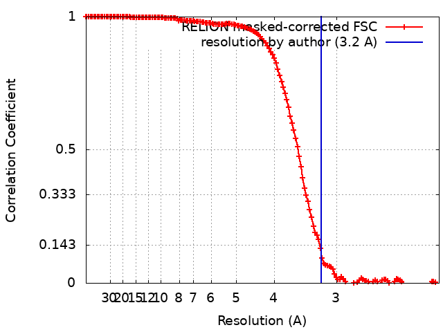
single particle reconstruction / cryo EM / Resolution: 3.2 Å
Journal: Nat Plants / Year: 2020 Title: Structure of a minimal photosystem I from the green alga Dunaliella salina. Authors: Annemarie Perez-Boerema / Daniel Klaiman / Ido Caspy / Sigal Y Netzer-El / Alexey Amunts / Nathan Nelson / Abstract: Solar energy harnessed by oxygenic photosynthesis supports most of the life forms on Earth. In eukaryotes, photosynthesis occurs in chloroplasts and is achieved by membrane-embedded macromolecular ...Solar energy harnessed by oxygenic photosynthesis supports most of the life forms on Earth. In eukaryotes, photosynthesis occurs in chloroplasts and is achieved by membrane-embedded macromolecular complexes that contain core and peripheral antennae with multiple pigments. The structure of photosystem I (PSI) comprises the core and light-harvesting (LHCI) complexes, which together form PSI-LHCI. Here we determined the structure of PSI-LHCI from the salt-tolerant green alga Dunaliella salina using X-ray crystallography and electron cryo-microscopy. Our results reveal a previously undescribed configuration of the PSI core. It is composed of only 7 subunits, compared with 14-16 subunits in plants and the alga Chlamydomonas reinhardtii, and forms the smallest known PSI. The LHCI is poorly conserved at the sequence level and binds to pigments that form new energy pathways, and the interactions between the individual Lhca1-4 proteins are weakened. Overall, the data indicate the PSI of D. salina represents a different type of the molecular organization that provides important information for reconstructing the plasticity and evolution of PSI.
History
Deposition
Apr 23, 2019
-
Header (metadata) release
Jun 12, 2019
-
Map release
Feb 19, 2020
-
Update
Oct 1, 2025
-
Current status
Oct 1, 2025
Processing site: PDBe / Status: Released
-
Structure visualization
Movie
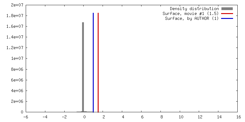
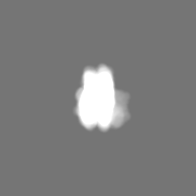
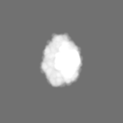
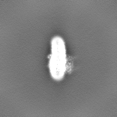
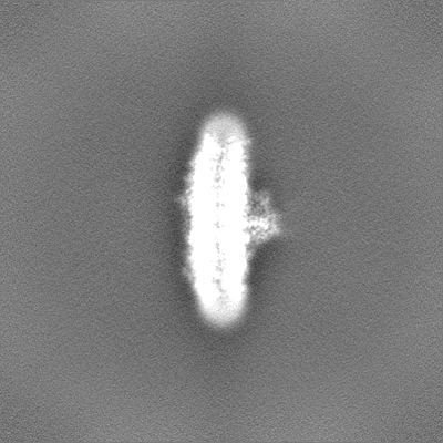
Surface view with section colored by density value
In the structure databanks used in Yorodumi, some data are registered as the other names, "COVID-19 virus" and "2019-nCoV". Here are the details of the virus and the list of structure data.
Jan 31, 2019. EMDB accession codes are about to change! (news from PDBe EMDB page)
EMDB accession codes are about to change! (news from PDBe EMDB page)
The allocation of 4 digits for EMDB accession codes will soon come to an end. Whilst these codes will remain in use, new EMDB accession codes will include an additional digit and will expand incrementally as the available range of codes is exhausted. The current 4-digit format prefixed with “EMD-” (i.e. EMD-XXXX) will advance to a 5-digit format (i.e. EMD-XXXXX), and so on. It is currently estimated that the 4-digit codes will be depleted around Spring 2019, at which point the 5-digit format will come into force.
The EM Navigator/Yorodumi systems omit the EMD- prefix.
Related info.:Q: What is EMD? / ID/Accession-code notation in Yorodumi/EM Navigator
Yorodumi is a browser for structure data from EMDB, PDB, SASBDB, etc.
This page is also the successor to EM Navigator detail page, and also detail information page/front-end page for Omokage search.
The word "yorodu" (or yorozu) is an old Japanese word meaning "ten thousand". "mi" (miru) is to see.
Related info.:EMDB / PDB / SASBDB / Comparison of 3 databanks / Yorodumi Search / Aug 31, 2016. New EM Navigator & Yorodumi / Yorodumi Papers / Jmol/JSmol / Function and homology information / Changes in new EM Navigator and Yorodumi
 Movie
Movie Controller


 Open data
 Basic information
 Map data
 Sample
 Keywords
 Function and homology information
 Dunaliella salina (plant)
 Authors
 Israel,
  Sweden, 3 items
  Citation
 Structure visualization
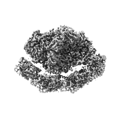
 Downloads & links
 emd_4883.png
 http://ftp.pdbj.org/pub/emdb/structures/EMD-4883


 Z (Sec.)
 Y (Row.)
 X (Col.)


 Sample components


 Processing
 Electron microscopy
 FIELD EMISSION GUN